Review
. 2011 May 16;24(5):618-39.
doi: 10.1021/tx200031q. Epub 2011 Apr 28.

Multifaceted roles of alkyltransferase and related proteins in DNA repair, DNA damage, resistance to chemotherapy, and research tools

Affiliations
Review

Multifaceted roles of alkyltransferase and related proteins in DNA repair, DNA damage, resistance to chemotherapy, and research tools

Anthony E Pegg. Chem Res Toxicol. .

Abstract

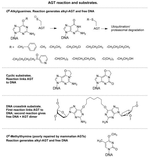
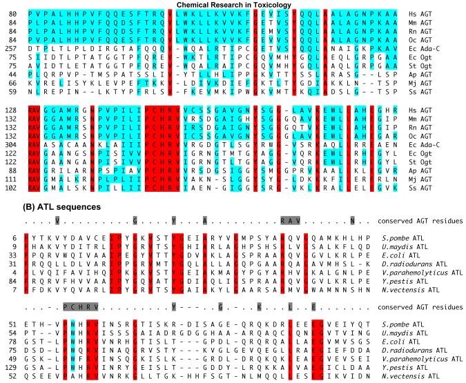
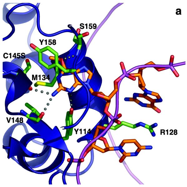
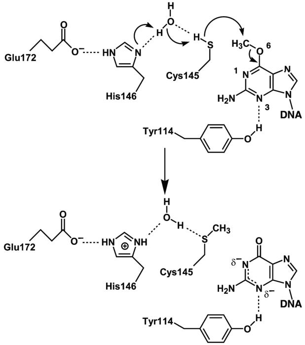
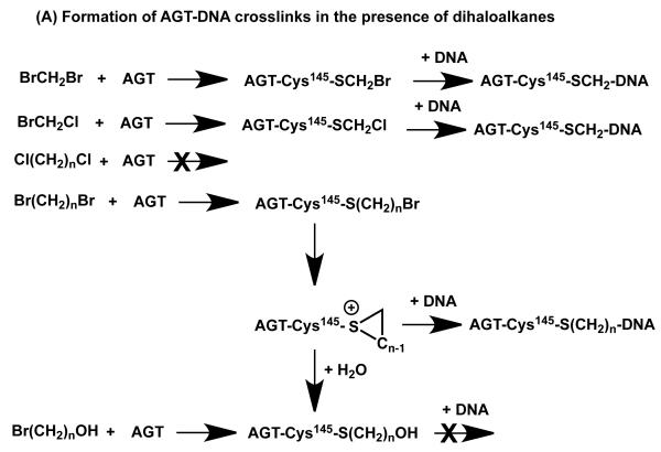
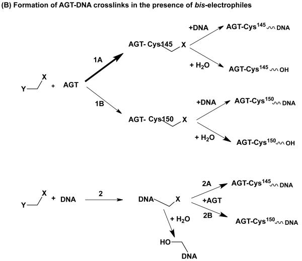
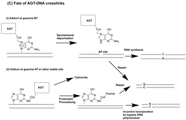
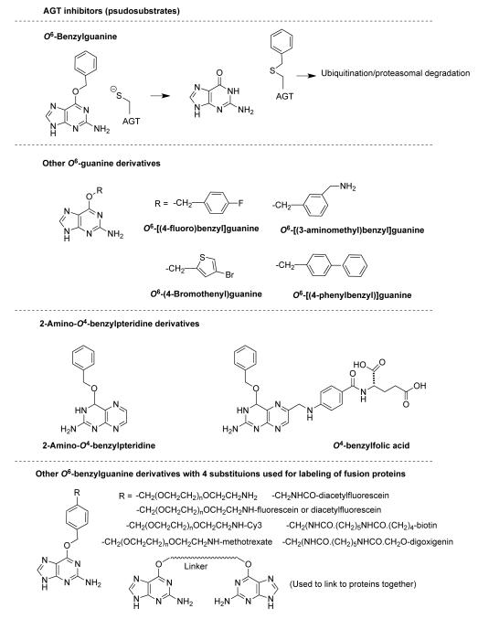
O(6)-Alkylguanine-DNA alkyltransferase (AGT) is a widely distributed, unique DNA repair protein that acts as a single agent to directly remove alkyl groups located on the O(6)-position of guanine from DNA restoring the DNA in one step. The protein acts only once, and its alkylated form is degraded rapidly. It is a major factor in counteracting the mutagenic, carcinogenic, and cytotoxic effects of agents that form such adducts including N-nitroso-compounds and a number of cancer chemotherapeutics. This review describes the structure, function, and mechanism of action of AGTs and of a family of related alkyltransferase-like proteins, which do not act alone to repair O(6)-alkylguanines in DNA but link repair to other pathways. The paradoxical ability of AGTs to stimulate the DNA-damaging ability of dihaloalkanes and other bis-electrophiles via the formation of AGT-DNA cross-links is also described. Other important properties of AGTs include the ability to provide resistance to cancer therapeutic alkylating agents, and the availability of AGT inhibitors such as O(6)-benzylguanine that might overcome this resistance is discussed. Finally, the properties of fusion proteins in which AGT sequences are linked to other proteins are outlined. Such proteins occur naturally, and synthetic variants engineered to react specifically with derivatives of O(6)-benzylguanine are the basis of a valuable research technique for tagging proteins with specific reagents.


Figures

Figure 1
Figure 1
AGT reaction and substrates
Figure 2
Figure 2
Key sequences in AGTs and ATLs. (A) shows the amino acid sequence alignment of the C-terminal domain containing the active site of AGT proteins from different species. The AGT sequences are from Homo sapiens (Hs), Mus musculus (Mm), Rattus norvegicus (Rn), Oryctolagus cuniculus (Oc), Escherichia coli (Ec), Salmonella typhimurium (St), Aeropyrum pernix (Ap), Methanococcus jannaschii (Mj) and Sulfolobus solfataricus (Ss). The amino acids that are conserved in the above AGTs are shown in red. The amino acids that similar or identical to human AGT protein are indicated in blue. The numbers on the left side of the sequences indicate the position of the amino acid in the AGT primary sequence. (B) shows amino acid sequence alignment of ATL proteins from different species and a comparison with conserved AGT residues. The ATL sequences are from Schizosaccharomyces pombe, Ustilago maydis, Escherichia coli, Deinococcus radiodurans, Vibrio parahaemolyticus, Yersinia pestis and Nematostella vectensis. The amino acids that are highly conserved in ATLs are indicated in red. The tryptophan residue that replaces the AGT alkyl acceptor site cysteine is indicated in blue. The amino acids that are highly conserved in AGTs from figure 1 are shaded grey and shown at the top of the aligned ATL sequences. The numbers on the left side of the sequences indicate the position of the amino acid in the primary sequence.
Figure 3
Figure 3
AGT:substrate complex and reaction mechanism. (A) Complex of C145S AGT mutant bound to DNA containing O6-methylguanine showing key residues Y114, R128, C145S and M134 in green. The DNA is shown in orange and the protein ribbon in blue. (B) Reaction mechanism of AGT showing activation of Cys 145 via interactions with Glu172-His146-water-C145. Redrawn with permission from refs (61, 62).
Figure 3
Figure 3
AGT:substrate complex and reaction mechanism. (A) Complex of C145S AGT mutant bound to DNA containing O6-methylguanine showing key residues Y114, R128, C145S and M134 in green. The DNA is shown in orange and the protein ribbon in blue. (B) Reaction mechanism of AGT showing activation of Cys 145 via interactions with Glu172-His146-water-C145. Redrawn with permission from refs (61, 62).
Figure 4
Figure 4
Formation of DNA-AGT crosslinks in the presence of dihaloalkanes and other bis-electrophiles. (A) Reactions of dihaloalkanes to generate crosslinks at Cys145. (B) Reactions of bis-electrophiles to generate crosslinks at Cys145 and Cys150. The chemical can react directly with AGT at either Cys145 or Cys150 as shown in 1A and 1B respectively. Further reaction with DNA then forms the AGT-DNA crosslink. The bis-electrophile may also react with DNA directly to generate a reactive species that can then react with AGT to produce the AGT-DNA crosslink at either Cys residue as shown in 2A and 2B. (C) The AGT-crosslinks can cause mutations or cytoxicity. (i) Attachment at the guanine N7 position generates an unstable linkage that can spontaneously depurinate to form an AP site which on DNA replication can then lead to the observed G:C to T:A transversions. (ii) More stable adducts, possibly at the guanine N2, although this has not been characterized, can be processed and copied by bypass polymerases to lead to the observed G:C to A:T transitions.
Figure 4
Figure 4
Formation of DNA-AGT crosslinks in the presence of dihaloalkanes and other bis-electrophiles. (A) Reactions of dihaloalkanes to generate crosslinks at Cys145. (B) Reactions of bis-electrophiles to generate crosslinks at Cys145 and Cys150. The chemical can react directly with AGT at either Cys145 or Cys150 as shown in 1A and 1B respectively. Further reaction with DNA then forms the AGT-DNA crosslink. The bis-electrophile may also react with DNA directly to generate a reactive species that can then react with AGT to produce the AGT-DNA crosslink at either Cys residue as shown in 2A and 2B. (C) The AGT-crosslinks can cause mutations or cytoxicity. (i) Attachment at the guanine N7 position generates an unstable linkage that can spontaneously depurinate to form an AP site which on DNA replication can then lead to the observed G:C to T:A transversions. (ii) More stable adducts, possibly at the guanine N2, although this has not been characterized, can be processed and copied by bypass polymerases to lead to the observed G:C to A:T transitions.
Figure 5
Figure 5
Inhibitors of AGT activity. References to the inhibitors and many related compounds are found in (112, 116, 165, 167, 170-174, 270). Although not shown here, additions to the N9 position are well tolerated and can be used to design additional inhibitors with improved solubility and other useful properties. The bottom section shows some of the compounds that can be used to label SNAP tags with useful chemical probes as described in section 10.1. Many additional compounds and variants of these probes have also been synthesized (254-262).
Figure 6
Figure 6
Properties of ATLs. Panel A shows overlay of the V. parahaemolyticus ATL (vpARL) structure (cyan) with the crystal structure of S. pombe ATL (spATL) bound to DNA containing O6-methylguanine (grey) The side chains of Tyr23, Arg37, and Trp54 are shown in red, blue, and yellow, respectively. Panels B-D show the inhibition of human AGT activity by ATLs and mutants at key residues. Panel B effect of vpAtl (triangles) and spAtl1 (circles). Panel C shows effect of mutating Tyr23 in vpAtl; wild type (black) triangles, Y23A (red triangles), and Y23F (red circles). D shows the effect of mutating Arg37 and Trp54 in vpAtl; wild type (black), R37A (blue) and W54A (gold). Reproduced from Figure 3 of reference (236).

References

    1. Pegg AE, Dolan ME, Moschel RC. Structure, function and inhibition of O6-alkylguanine-DNA alkyltransferase. Progr. Nucleic Acids Res. Mol. Biol. 1995;51:167–223. - PubMed
    1. Pegg AE. Repair of O6-alkylguanine by alkyltransferases. Mutat. Res. 2000;462:83–100. - PubMed
    1. Margison G, Povey AC, Kaina B, Santibáñez-Koref MF. Variability and regulation of O6-alkylguanine-DNA alkyltransferase. Carcinogenesis. 2003;24:625–635. - PubMed
    1. Gerson SL. MGMT: its role in cancer aetiology and cancer therapeutics. Nat. Rev. Cancer. 2004;4:296–307. - PubMed
    1. Mishina Y, Duguid EM, He C. Direct reversal of DNA alkylation damage. Chem. Rev. 2006;106:215–232. - PMC - PubMed

Publication types

Substances