

In vitro functional testing of endothelial progenitor cells that overexpress thrombomodulin
- PMID: 21466416
- PMCID: PMC3142633
- DOI: 10.1089/ten.TEA.2010.0631
In vitro functional testing of endothelial progenitor cells that overexpress thrombomodulin
Abstract
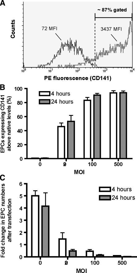
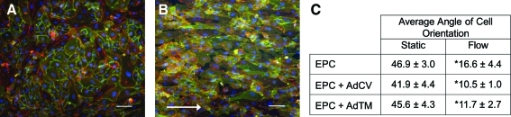
This study investigated the augmentation of endothelial progenitor cell (EPC) thromboresistance by using gene therapy to overexpress thrombomodulin (TM), an endothelial cell membrane glycoprotein that has potent anti-coagulant properties. Late outgrowth EPCs were isolated from peripheral blood of patients with documented coronary artery disease and transfected with an adenoviral vector containing human TM. EPC transfection conditions for maximizing TM expression, transfection efficiency, and cell viability were employed. TM-overexpressing EPCs had a fivefold increase in the rate of activated protein C production over native EPCs and EPCs transfected with an adenoviral control vector expressing β-galactosidase (p<0.05). TM upregulation caused a significant threefold reduction in platelet adhesion compared to native EPCs, and a 12-fold reduction compared to collagen I-coated wells. Additionally, the clotting time of TM-transfected EPCs incubated with whole blood was significantly extended by 19% over native cells (p<0.05). These data indicate that TM-overexpression has the potential to improve the antithrombotic performance of patient-derived EPCs for endothelialization applications.
Figures


Similar articles
-
Patient-derived endothelial progenitor cells improve vascular graft patency in a rodent model.Acta Biomater. 2012 Jan;8(1):201-8. doi: 10.1016/j.actbio.2011.09.002. Epub 2011 Sep 6. Acta Biomater. 2012. PMID: 21945828 Free PMC article.
-
Genetic engineering with endothelial nitric oxide synthase improves functional properties of endothelial progenitor cells from patients with coronary artery disease: an in vitro study.Basic Res Cardiol. 2009 Nov;104(6):739-49. doi: 10.1007/s00395-009-0039-x. Epub 2009 May 29. Basic Res Cardiol. 2009. PMID: 19479297
-
CD133+ endothelial progenitor cells as a potential cell source for a bioartificial glomerulus.Tissue Eng Part A. 2009 Oct;15(10):3173-82. doi: 10.1089/ten.TEA.2009.0050. Tissue Eng Part A. 2009. PMID: 19358628
-
The biocompatibility of titanium cardiovascular devices seeded with autologous blood-derived endothelial progenitor cells: EPC-seeded antithrombotic Ti implants.Biomaterials. 2011 Jan;32(1):10-8. doi: 10.1016/j.biomaterials.2010.08.073. Epub 2010 Nov 5. Biomaterials. 2011. PMID: 20926131 Free PMC article.
-
Tissue engineering of bladder using vascular endothelial growth factor gene-modified endothelial progenitor cells.Int J Artif Organs. 2011 Dec;34(12):1137-46. doi: 10.5301/ijao.5000069. Int J Artif Organs. 2011. PMID: 22198599
Cited by
-
Nanoengineered injectable hydrogels for wound healing application.Acta Biomater. 2018 Apr 1;70:35-47. doi: 10.1016/j.actbio.2018.01.045. Epub 2018 Feb 7. Acta Biomater. 2018. PMID: 29425720 Free PMC article.
-
Microfluidic techniques for development of 3D vascularized tissue.Biomaterials. 2014 Aug;35(26):7308-25. doi: 10.1016/j.biomaterials.2014.04.091. Epub 2014 Jun 3. Biomaterials. 2014. PMID: 24906345 Free PMC article. Review.
-
Patient-derived endothelial progenitor cells improve vascular graft patency in a rodent model.Acta Biomater. 2012 Jan;8(1):201-8. doi: 10.1016/j.actbio.2011.09.002. Epub 2011 Sep 6. Acta Biomater. 2012. PMID: 21945828 Free PMC article.
-
In vivo assessment of two endothelialization approaches on bioprosthetic valves for the treatment of chronic deep venous insufficiency.J Biomed Mater Res B Appl Biomater. 2016 Nov;104(8):1610-1621. doi: 10.1002/jbm.b.33507. Epub 2015 Aug 28. J Biomed Mater Res B Appl Biomater. 2016. PMID: 26316151 Free PMC article.
-
Endothelial outgrowth cells: function and performance in vascular grafts.Tissue Eng Part B Rev. 2014 Aug;20(4):294-303. doi: 10.1089/ten.TEB.2013.0285. Epub 2013 Oct 10. Tissue Eng Part B Rev. 2014. PMID: 24004404 Free PMC article. Review.
References
-
- Meinhart J.G. Deutsch M. Fischlein T. Howanietz N. Fröschl A. Zilla P. Clinical autologous in vitro endothelialization of 153 infrainguinal ePTFE grafts. Ann Thorac Surg. 2001;71:S327. - PubMed
-
- Scott-Burden T. Tock C.L. Bosely J.P. Clubb F.J., Jr. Parnis S.M. Schwarz J.J., et al. Nonthrombogenic, adhesive cellular lining for left ventricular assist devices. Circulation. 1998;98:II339. - PubMed
-
- Schmidt D. Mol A. Breymann C. Achermann J. Odermatt B. Gossi M., et al. Living autologous heart valves engineered from human prenatally harvested progenitors. Circulation. 2006;114:I-125. - PubMed
-
- Kannan R.Y. Salacinski H.J. Butler P.E. Hamilton G. Seifalian A.M. Current status of prosthetic bypass grafts: a review. J Biomed Mater Res B Appl Biomater. 2005;74:570. - PubMed
Publication types
MeSH terms
Substances
Grants and funding
LinkOut - more resources
Full Text Sources
Medical
