. 2011 Jul;133(3):288-95.
doi: 10.1111/j.1365-2567.2011.03437.x. Epub 2011 Apr 6.

The stromal cell antigen CD248 (endosialin) is expressed on naive CD8+ human T cells and regulates proliferation

Affiliations

The stromal cell antigen CD248 (endosialin) is expressed on naive CD8+ human T cells and regulates proliferation

Debbie L Hardie et al. Immunology. 2011 Jul.

Abstract

CD248 (endosialin) is a transmembrane glycoprotein that is dynamically expressed on pericytes and fibroblasts during tissue development, tumour neovascularization and inflammation. Its role in tissue remodelling is associated with increased stromal cell proliferation and migration. We show that CD248 is also uniquely expressed by human, but not mouse (C57BL/6), CD8(+) naive T cells. CD248 is found only on CD8(+) CCR7(+) CD11a(low) naive T cells and on CD8 single-positive T cells in the thymus. Transfection of the CD248 negative T-cell line MOLT-4 with CD248 cDNA surprisingly reduced cell proliferation. Knock-down of CD248 on naive CD8 T cells increased cell proliferation. These data demonstrate opposing functions for CD248 on haematopoietic (CD8(+)) versus stromal cells and suggests that CD248 helps to maintain naive CD8(+) human T cells in a quiescent state.


Figures

Figure 1
Figure 1
CD248 is expressed on human stromal cells and cells with lymphoid morphology. Examples of CD248+ lymphoid cells that reside in the extrafollicular area (EFA) but not the germinal centre (GC) are circled. These are CD3+ cells (a) that are CD4 CD8+ and were present in tonsil (b), spleen (c) and thymus (d). Boxes indicate areas shown at higher magnification on the right. Data are representative images from at least three different donors for tonsil and spleen and one for thymus. Scale bars represent 50 μm.
Figure 2
Figure 2
CD248+ T cells are not present in C57/BL6 mice. CD248+ (green) T cells (CD3+ red) were not present in lymph node (a), spleen (b) or thymus (c) of mice. RP, red pulp; WP, white pulp. Scale bars represent 50 μm. Boxes indicate areas shown at higher magnification on the right. Data are representative images from at least three different mice.
Figure 3
Figure 3
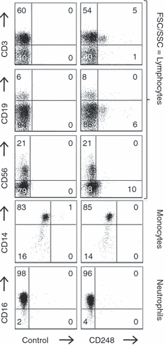
CD248 is expressed on a subset of CD3+ T cells from human blood. Flow cytometry of CD248 antibody B1/35.1 in comparison with negative controls showing that CD248 is expressed on a subset (8·5%) of CD3+ lymphocytes from blood. CD19+, CD56+, CD14+ and CD16+ cells are CD248. B, T and natural killer cells are forward scatter/side scatter (FSC/SSC) gated for lymphocytes, CD14+ cells are FSC/SSC gated for monocytes and CD16+ cells are FSC/SSC gated for neutrophils. Data are representative plots from a minimum of three experiments from different donors.
Figure 4
Figure 4
CD248 is expressed on naive CD8+ T cells and mature human thymocytes. CD248 is expressed by a distinct subset of CD8+ CD3+ T cells that is CD45RA+ CCR7+ CD11alow and is present in blood and tonsil (a). Samples are gated in comparison with isotype controls. Data are representative plots from a minimum of five samples each. (b) Western blot (n = 1) showing CD248 protein expression only in rheumatoid arthritis synovial fibroblasts and CD8+ CD45RA+ T cells and not other T-cell subsets or HUVEC. (c) CD248+ CD3+ thymocytes (n = 1) are only found in the single-positive CD4 CD8+ population of T cells.
Figure 5
Figure 5
Human (hu) CD248 transfection of MOLT-4 T cells resulted in decreased proliferation. CD248 monoclonal antibody B1/35.1 staining of surface (a) and fixed (b) MOLT-4 T cells transfected with either huCD248-pcDNA3.1 or pcDNA3.1 vector alone show that only the CD248 transfected line has high expression of CD248. Insets in (b) are nuclear counterstains. Scale bars represent 10 μm (n≥ 3). Comparison of the [3H]thymidine uptake of MOLT-4 T cells transfected with CD248-pcDNA3.1 (•) or pcDNA3.1 vector alone (○) shows that CD248+ cells have lower spontaneous proliferative capacity than their vector alone-transfected counterparts (c). This was demonstrated across a range of cell numbers and on day 1 of culture (n = 7). *P < 0·05, **P < 0·01, ***P < 0·001. The Student's paired t-test was applied to compare the statistical significance of linked data. Error bars represent SEM.
Figure 6
Figure 6
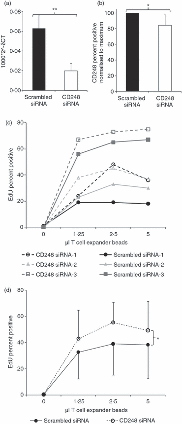
CD248 expression is associated with decreased proliferation of naive CD8 T cells. Quantitative PCR was used to demonstrate significantly lower levels (**P < 0·01) of CD248 RNA expression in CD248 small interfering (si) RNA-treated cells compared with controls (n = 3) (a). Significantly reduced protein expression (*P < 0·05) of CD248 in CD248 siRNA-treated cells compared with controls was also demonstrated (n = 3) (b). Cells were cultured with 0, 1·25, 2·5 or 5 μl T-cell expander beads for 3 days then their proliferation was compared using an 5-ethynyl-2′-deoxyuridine (EdU) incorporation assay. CD248 siRNA-treated naive CD8 T cells (formula image) exhibit higher EdU incorporation than scrambled siRNA-treated cells (formula image). Data from three different donors are shown in (c) and the data are grouped in (d) showing CD248 siRNA treated naive CD8 T cells (formula image) exhibit significantly higher (*P < 0·05) EdU incorporation than scrambled siRNA-treated cells (formula image). The Student's paired t-test was applied to compare the statistical significance of linked data. Error bars represent SEM.

References

    1. Rettig WJ, Garin-Chesa P, Healey JH, Su SL, Jaffe EA, Old LJ. Identification of endosialin, a cell surface glycoprotein of vascular endothelial cells in human cancer. Proc Natl Acad Sci USA. 1992;89:10832–6. - PMC - PubMed
    1. Christian S, Ahorn H, Koehler A, et al. Molecular cloning and characterization of endosialin, a C-type lectin-like cell surface receptor of tumor endothelium. J Biol Chem. 2001;276:7408–14. - PubMed
    1. Greenlee MC, Sullivan SA, Bohlson SS. CD93 and related family members: their role in innate immunity. Curr Drug Targets. 2008;9:130–8. - PubMed
    1. St Croix B, Rago C, Velculescu V, et al. Genes expressed in human tumor endothelium. Science. 2000;289:1197–202. - PubMed
    1. Bagley RG, Honma N, Weber W, et al. Endosialin/TEM 1/CD248 is a pericyte marker of embryonic and tumor neovascularization. Microvasc Res. 2008;76:180–8. - PubMed

Publication types