

Computed tomography measurements of different dimensions of maxillary and frontal sinuses
- PMID: 21466703
- PMCID: PMC3080316
- DOI: 10.1186/1471-2342-11-8
Computed tomography measurements of different dimensions of maxillary and frontal sinuses
Abstract
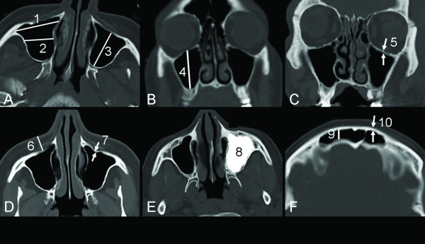
Background: We have previously proposed the use of Doppler ultrasound to non-invasively stage sinus infection, as we showed that acoustic streaming could be generated in nonpurulent sinus secretions and helped to distinguish it from mucopurulent sinus secretions. In order to continue this development of a clinically applicable Doppler equipment, we need to determine different dimensions of the paranasal sinuses, especially the thickness of the anterior wall of the maxillary sinus (at the canine fossa). To the best of our knowledge, this is the first report on the thickness of the canine fossa. This study aimed to (a) estimate different dimensions of the maxillary and frontal sinuses measured on computed tomography (CT) of the head, (b) define cut-off values for the normal upper and lower limits of the different measured structures, (c) determine differences in age, side and gender, (d) compare manually and automatically estimated maxillary sinuses volumes, and (e) present incidental findings in the paranasal sinuses among the study patients.
Methods: Dimensions of 120 maxillary and frontal sinuses from head CTs were measured independently by two radiologists.
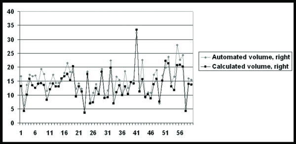
Results: The mean value of the maxillary sinus volume was 15.7±5.3 cm3 and significantly larger in males than in females (P=0.004). There was no statistically significant correlation between the volume of maxillary sinuses with age or side. The mean value of the bone thickness at the canine fossa was 1.1±0.4 mm. The automatically estimated volume of the maxillary sinuses was 14-17% higher than the calculated volume. There was high interobserver agreement with regard to the different measurements performed in this study. Different types of incidental findings of the paranasal sinuses were found in 35% of the patients.
Conclusion: We presented different dimensions of the maxillary and frontal sinuses on CTs. We believe that our data are necessary for further development of a clinically applicable Doppler equipment for staging rhinosinusitis.
Figures
References
-
- Kawarai Y, Fukushima K, Ogawa T, Nishizaki K, Gunduz M, Fujimoto M, Masuda Y. Volume quantification of healthy paranasal cavity by three-dimensional CT imaging. Acta Otolaryngol Suppl. 1999;540:45–49. - PubMed
Publication types
MeSH terms
LinkOut - more resources
Full Text Sources
Other Literature Sources
Medical
Research Materials
