. 2010:75:403-18.
doi: 10.1101/sqb.2010.75.038. Epub 2011 Apr 5.

Centromere identity, function, and epigenetic propagation across cell divisions

Affiliations

Centromere identity, function, and epigenetic propagation across cell divisions

B E Black et al. Cold Spring Harb Symp Quant Biol. 2010.

Abstract

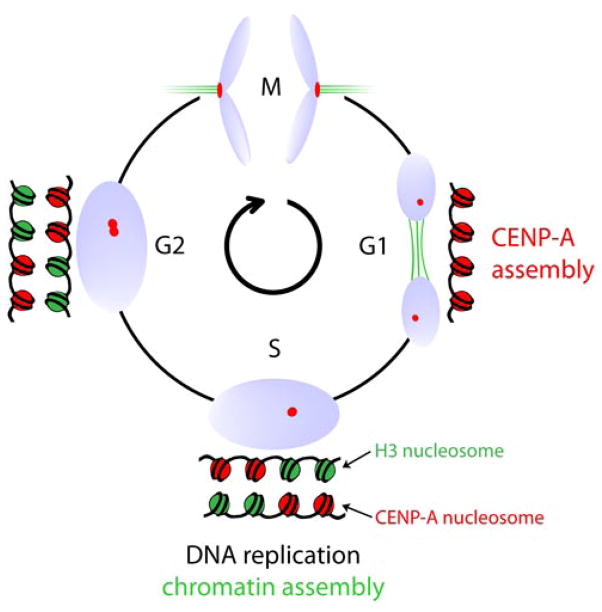
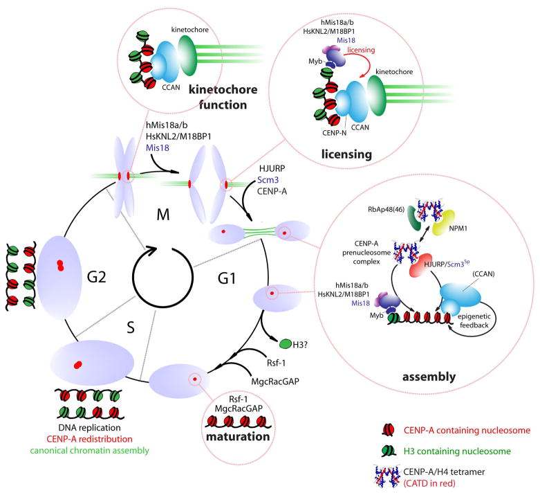
The key to understanding centromere identity is likely to lie in the chromatin containing the histone H3 variant CENP-A. CENP-A is the prime candidate to carry the epigenetic information that specifies the chromosomal location of the centromere in nearly all eukaryotic species, raising questions fundamental to understanding chromosome inheritance: How is the epigenetic centromere mark propagated? What physical properties of CENP-A-containing complexes are important for epigenetically marking centromeres? What are the molecules that recognize centromeric chromatin and serve as the foundation for the mitotic kinetochore? We discuss recent advances from our research groups that have yielded substantial insight into these questions and present our current understanding of the centromere. Future work promises an understanding of the molecular processes that confer fidelity to genome transmission at cell division.


Figures

Figure 1
Figure 1
Epigenetic centromere specification. (A) DNA at normal human centromeres is repetitive with a monomer length of ~171 bp multimerized for megabase stretches. While this general theme in centromere organization is seen in most eukaryotes, centromeric DNA sequences are amongst some of the most rapidly evolving sequences in the genome. (B) Stable inheritance of a human neocentromere after centromere relocation along an intact human chromosome 4 (Amor et al. 2004). At left is the family pedigree, showing the generational inheritance of a neocentromere containing variant chromosome 4 (‘PD-NC4’; black bar). The chromosomal allele containing the neocentromere was inherited from the paternal grandfather of the brother and sister who initially were found to carry PD-NC4. The neocentromere is carried by their father, and the grandfather was not available for study. (C) Relocation of both centromere-specifying chromatin and inner centromere components to the PD-NC4 neocentromere(Bassett et al. 2010). Anti-centromere antisera (ACA) recognizes both CENP-A at the neocentromere (arrowhead) and CENP-B at the silenced centromere at the original location (asterisk). Kinetochore-forming components, represented by the CENP-A-binding protein, CENP-C, vacate the original site and relocate to the neocentromere (Amor et al. 2004). Inner centromere components, represented by the Aurora B kinase also vacate the original site and relocate to chromosome arm positions proximal to the neocentromere(Bassett et al. 2010).
Figure 2
Figure 2
Initial evidence linking the targeting of CENP-A to centromeres with physical divergence from conventional histone H3 (Black et al. 2004). (A) Diagram of a histone H3 chimera containing the CENP-A targeting domain (CATD). (B) Centromere targeting of H3CATD. (C) Hydrogen/deuterium exchange profile of histone H4 bound to conventional H3, the H3CATD chimera, and CENP-A. The nearly identically localized blue regions on histone H4 within (CENP-A:H4)2 and (H3CATD:H4)2 heterotetramers are >10-fold slower to exchange amide protons with deuterons in heavy water than from any portion within conventional (H3:H4)2, indicating substantial rigidity imparted by the CATD.
Figure 3
Figure 3
(CENP-A/H4)2 heterotetramers structurally deviate from their conventional counterparts. (A) Crystal structure of the (CENP-A:H4)2 heterotetramer (PDB ID 3NQJ)(Sekulic et al. 2010) highlighting features that distinguish it from conventional (H3:H4)2. (B) Surface alterations encoded by the CATD of CENP-A include a basic charged protrusion (circled) that is bulged further away from the helical core of the complex and of the opposite charge as on the counterpart (H3:H4)2 heterotetramer. (C) Solution measurements of (CENP-A:H4)2 indicate that it is compacted by ~10 Å relative to its conventional counterpart, as predicted by the compact structure of (CENP-A:H4)2 in crystal form. The mesh indicates the molecular envelope corresponding to the rotational state that best matches the SAXS data collected for each type of heterotetramer.
Figure 4
Figure 4
A pool of CENP-A-SNAP, synthesized during S phase, was fluorescently pulse labeled in G2 phase. Cells were then cycled through mitosis and fixed. Accumulation of nascent CENP-A-SNAP (pulse labeled SNAP, red) at centromeres is evident in cells in late telophase, marked by reformed nuclei (DNA, Blue) and mid-bodies identifying daughter cells (microtubules, green).
Figure 5
Figure 5
A schematic representation of the temporal uncoupling of DNA replication and canonical chromatin assembly in S phase from centromeric nucleosome replication in G1.
Figure 6
Figure 6
Identification of centromere constituents. (A) Tandem affinity purification was used to identify the set of centromere proteins most closely associated with the CENP-A nucleosome (CENP-ANAC) by affinity purification of intact CENP-A nucleosomes derived from an MNase digested chromatin fraction. (B) Serial affinity purification of newly identified CENP-ANAC proteins was used to identify the more distal constitutive components of the centromere that were not in close proximity to the CENP-A nucleosome. (C) Cartoon model of centromere organization from CENP-A nucleosomes to outer kinetochore formation (Foltz et al. 2006).
Figure 7
Figure 7
Isolation of pre-nucleosomal histone complexes. (A) Affinity purifications of CENP-A, H3CATD and histone H3.1 were conducted from chromatin-free extracts to identify CENP-A specific preassembly complexes (Foltz et al. 2009). Prenucleosomal CENP-A is uniquely associated with HJURP and NPM1 compared with histone H3.1. The CATD domain of CENP-A mediates the recruitment of HJURP as the H3CATD chimeric protein, which is recruited to centromeres, interacts with HJURP. (B) Model of the distinct histone chaperone complexes utilized by the histone H3 variants.
Figure 8
Figure 8
CENP-A assembles into octameric nucleosomes with conventional handedness of DNA wrapping (Sekulic et al. 2010). (A) Histone content of assembled H3- and CENP-A-containing nucleosomes. (B) Digestion of nucleosome arrays with micrococcal nuclease reveals that both H3-and CENP-A-containing nucleosomes protect ~150 bp of DNA. (C–F) Topological analysis of H3-(C and D) and CENP-A-containing (E and F) nucleosomes. Analysis by gel electrophoresis in the absence (C and E) or presence (D and F) of chloroquine reveals that both H3- and CENP-A-containing nucleosomes wrap DNA in a left-handed manner.
Figure 9
Figure 9
Model representing the centromeric chromatin cycle. CENP-A-containing nucleosomes are redistributed during DNA replication in S phase. The resulting mixed CENP-A/H3 chromatin supports kinetochore formation during mitotis. An unknown mitotic signal triggers “licensing” of centromeres by Mis18:KNL2 proteins in anaphase for subsequence assembly of CENP-A. The molecular nature of the licensing step is unknown but is likely precedes recruitment of CENP-A as a separate step. Assembly of new CENP-A is mediated by the HJURP-containing prenucleosomal complex throughout the first hours of G1. Targeting of the prenucleosomal complex may involve the Mis18:KNL2 proteins or CCAN members. Assembly of CENP-A is predicted to involve exchange of H3 from centromeric chromatin and may require additional activities provided by the RSF complex and the small GTPase activating protein MgcRacGAP in late G1 (provisionally termed ‘maturation’).

References

    1. Ahmad K, Henikoff S. Histone H3 variants specify modes of chromatin assembly. Proc Natl Acad Sci U S A. 2002a;99(Suppl 4):16477–16484. - PMC - PubMed
    1. Ahmad K, Henikoff S. The histone variant H3.3 marks active chromatin by replication-independent nucleosome assembly. Mol Cell. 2002b;9(6):1191–1200. - PubMed
    1. Allshire RC, Karpen GH. Epigenetic regulation of centromeric chromatin: old dogs, new tricks? Nat Rev Genet. 2008;9(12):923–937. - PMC - PubMed
    1. Amano M, Suzuki A, Hori T, Backer C, Okawa K, Cheeseman IM, Fukagawa T. The CENP-S complex is essential for the stable assembly of outer kinetochore structure. J Cell Biol. 2009;186(2):173–182. - PMC - PubMed
    1. Amor DJ, Bentley K, Ryan J, Perry J, Wong L, Slater H, Choo KH. Human centromere repositioning “in progress”. Proc Natl Acad Sci U S A. 2004;101(17):6542–6547. - PMC - PubMed

Publication types

LinkOut - more resources