Analysis of two in planta expressed LysM effector homologs from the fungus Mycosphaerella graminicola reveals novel functional properties and varying contributions to virulence on wheat
- PMID: 21467214
- PMCID: PMC3177273
- DOI: 10.1104/pp.111.176347
Analysis of two in planta expressed LysM effector homologs from the fungus Mycosphaerella graminicola reveals novel functional properties and varying contributions to virulence on wheat
Abstract
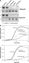
Secreted effector proteins enable plant pathogenic fungi to manipulate host defenses for successful infection. Mycosphaerella graminicola causes Septoria tritici blotch disease of wheat (Triticum aestivum) leaves. Leaf infection involves a long (approximately 7 d) period of symptomless intercellular colonization prior to the appearance of necrotic disease lesions. Therefore, M. graminicola is considered as a hemibiotrophic (or necrotrophic) pathogen. Here, we describe the molecular and functional characterization of M. graminicola homologs of Ecp6 (for extracellular protein 6), the Lysin (LysM) domain-containing effector from the biotrophic tomato (Solanum lycopersicum) leaf mold fungus Cladosporium fulvum, which interferes with chitin-triggered immunity in plants. Three LysM effector homologs are present in the M. graminicola genome, referred to as Mg3LysM, Mg1LysM, and MgxLysM. Mg3LysM and Mg1LysM genes were strongly transcriptionally up-regulated specifically during symptomless leaf infection. Both proteins bind chitin; however, only Mg3LysM blocked the elicitation of chitin-induced plant defenses. In contrast to C. fulvum Ecp6, both Mg1LysM and Mg3LysM also protected fungal hyphae against plant-derived hydrolytic enzymes, and both genes show significantly more nucleotide polymorphism giving rise to nonsynonymous amino acid changes. While Mg1LysM deletion mutant strains of M. graminicola were fully pathogenic toward wheat leaves, Mg3LysM mutant strains were severely impaired in leaf colonization, did not trigger lesion formation, and were unable to undergo asexual sporulation. This virulence defect correlated with more rapid and pronounced expression of wheat defense genes during the symptomless phase of leaf colonization. These data highlight different functions for MgLysM effector homologs during plant infection, including novel activities that distinguish these proteins from C. fulvum Ecp6.
Figures


Similar articles
-
Mycosphaerella graminicola LysM effector-mediated stealth pathogenesis subverts recognition through both CERK1 and CEBiP homologues in wheat.Mol Plant Microbe Interact. 2014 Mar;27(3):236-43. doi: 10.1094/MPMI-07-13-0201-R. Mol Plant Microbe Interact. 2014. PMID: 24073880
-
Three LysM effectors of Zymoseptoria tritici collectively disarm chitin-triggered plant immunity.Mol Plant Pathol. 2021 Jun;22(6):683-693. doi: 10.1111/mpp.13055. Epub 2021 Apr 1. Mol Plant Pathol. 2021. PMID: 33797163 Free PMC article.
-
A secreted LysM effector protects fungal hyphae through chitin-dependent homodimer polymerization.PLoS Pathog. 2020 Jun 23;16(6):e1008652. doi: 10.1371/journal.ppat.1008652. eCollection 2020 Jun. PLoS Pathog. 2020. PMID: 32574207 Free PMC article.
-
Previous bottlenecks and future solutions to dissecting the Zymoseptoria tritici-wheat host-pathogen interaction.Fungal Genet Biol. 2015 Jun;79:24-8. doi: 10.1016/j.fgb.2015.04.005. Fungal Genet Biol. 2015. PMID: 26092786 Free PMC article. Review.
-
Dissecting the Molecular Interactions between Wheat and the Fungal Pathogen Zymoseptoria tritici.Front Plant Sci. 2016 Apr 15;7:508. doi: 10.3389/fpls.2016.00508. eCollection 2016. Front Plant Sci. 2016. PMID: 27148331 Free PMC article. Review.
Cited by
-
A conserved proline residue in Dothideomycete Avr4 effector proteins is required to trigger a Cf-4-dependent hypersensitive response.Mol Plant Pathol. 2016 Jan;17(1):84-95. doi: 10.1111/mpp.12265. Epub 2015 May 7. Mol Plant Pathol. 2016. PMID: 25845605 Free PMC article.
-
Zymoseptoria tritici white-collar complex integrates light, temperature and plant cues to initiate dimorphism and pathogenesis.Nat Commun. 2022 Sep 26;13(1):5625. doi: 10.1038/s41467-022-33183-2. Nat Commun. 2022. PMID: 36163135 Free PMC article.
-
Crosstalk of Signaling Mechanisms Involved in Host Defense and Symbiosis Against Microorganisms in Rice.Curr Genomics. 2016 Aug;17(4):297-307. doi: 10.2174/1389202917666160331201602. Curr Genomics. 2016. PMID: 27499679 Free PMC article.
-
LysM receptors in Coffea arabica: Identification, characterization, and gene expression in response to Hemileia vastatrix.PLoS One. 2022 Feb 10;17(2):e0258838. doi: 10.1371/journal.pone.0258838. eCollection 2022. PLoS One. 2022. PMID: 35143519 Free PMC article.
-
Predication of the Effector Proteins Secreted by Fusarium sacchari Using Genomic Analysis and Heterogenous Expression.J Fungi (Basel). 2022 Jan 6;8(1):59. doi: 10.3390/jof8010059. J Fungi (Basel). 2022. PMID: 35049998 Free PMC article.
References
-
- Adhikari TB, Balaji B, Breeden J, Goodwin SB. (2007) Resistance of wheat to Mycosphaerella graminicola involves early and late peaks of gene expression. Physiol Mol Plant Pathol 71: 55–68
-
- An G, Evert PR, Mitra A, Ha SB. (1988) Binary vectors. Gelvin SB, Schilperroort RA, eds, Plant Molecular Biology Manual A3. Kluwer Academic Press, Dordrecht, The Netherlands, pp 1–19
-
- Barber MS, Bertram RE, Ride JP. (1989) Chitin oligosaccharides elicit lignification in wounded wheat leaves. Physiol Mol Plant Pathol 34: 3–12
-
- Bielawski JP, Yang Z. (2005) Maximum likelihood methods for detecting adaptive protein evolution. Nielsen R, ed, Statistical Methods in Molecular Evolution. Springer, New York, pp 103–124
Publication types
MeSH terms
Substances
Grants and funding
LinkOut - more resources
Full Text Sources
Other Literature Sources

