

HLA-DR-presented peptide repertoires derived from human monocyte-derived dendritic cells pulsed with blood coagulation factor VIII
- PMID: 21467215
- PMCID: PMC3108829
- DOI: 10.1074/mcp.M110.002246
HLA-DR-presented peptide repertoires derived from human monocyte-derived dendritic cells pulsed with blood coagulation factor VIII
Abstract
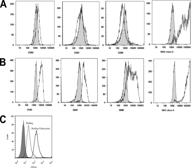
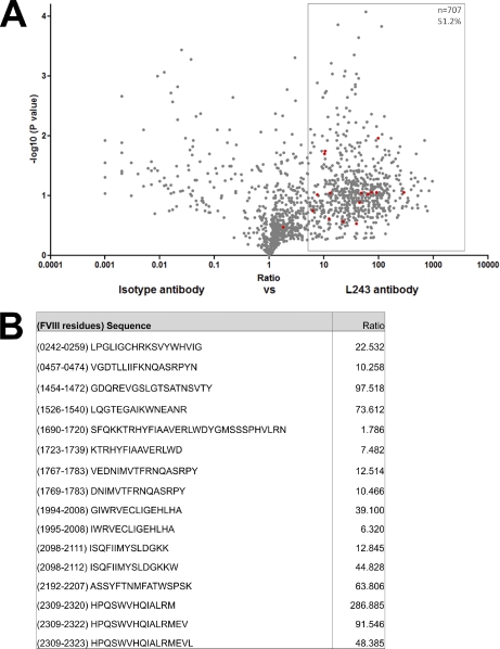
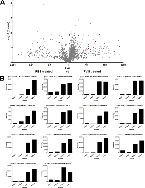
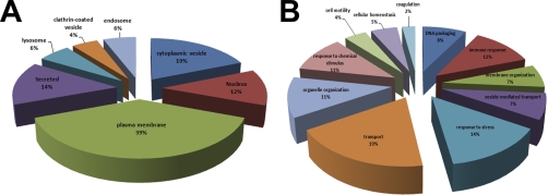
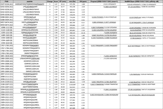
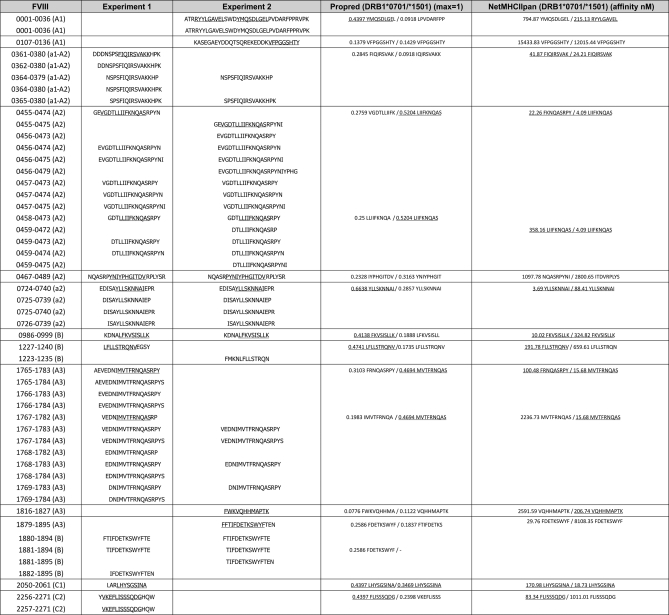
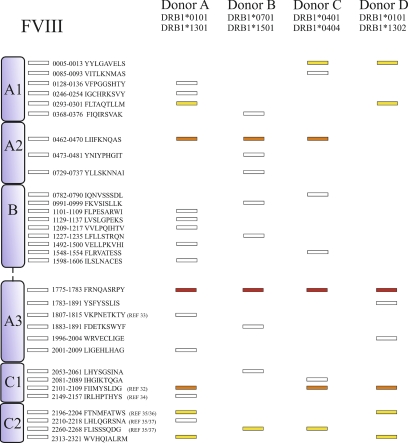
Activation of T-helper cells is dependent upon the appropriate presentation of antigen-derived peptides on MHC class II molecules expressed on antigen presenting cells. In the current study we explored the repertoire of peptides presented on MHC class II molecules on human monocyte derived dendritic cells (moDCs) from four HLA-typed healthy donors. MHC class II-bound peptides could be routinely recovered from small cultures containing 5 × 10(6) cells. A fraction of the identified peptides were derived from proteins localized in the plasma membrane, endosomes, and lysosomes, but the majority of peptides that were presented on MHC class II originate from other organelles. Subsequently, we studied the antigen-specific peptide repertoire after endocytosis of a soluble antigen. Blood coagulation factor VIII (FVIII) was chosen as the antigen since our current knowledge on MHC class II presented peptides derived from this immunogenic therapeutic protein is limited. Analysis of the total repertoire of MHC class II-associated peptides revealed that per individual sample 20-50 FVIII-derived peptides were presented on FVIII-pulsed moDCs. Repertoires of FVIII-derived peptides eluted from moDCs derived from a panel of four HLA typed donors revealed that some MHC class II-presented FVIII peptides were presented by multiple donors, whereas the presentation of other FVIII peptides was donor-specific. In total 32 different core peptides were presented on FVIII-pulsed moDCs from four HLA-typed donors. Together our findings provide an unbiased approach to identify peptides that are presented by MHC class II on antigen-loaded moDCs from individual donors.
Figures
Similar articles
-
Limited promiscuity of HLA-DRB1 presented peptides derived of blood coagulation factor VIII.PLoS One. 2013 Nov 14;8(11):e80239. doi: 10.1371/journal.pone.0080239. eCollection 2013. PLoS One. 2013. PMID: 24244658 Free PMC article.
-
FVIII peptides presented on HLA-DP and identification of an A3 domain peptide binding with high affinity to the commonly expressed HLA-DP4.Haematologica. 2025 Jun 1;110(6):1316-1327. doi: 10.3324/haematol.2024.286204. Epub 2024 Dec 12. Haematologica. 2025. PMID: 39665218 Free PMC article.
-
Comparative profiling of HLA-DR and HLA-DQ associated factor VIII peptides presented by monocyte-derived dendritic cells.Haematologica. 2018 Jan;103(1):172-178. doi: 10.3324/haematol.2017.175083. Epub 2017 Oct 12. Haematologica. 2018. PMID: 29025906 Free PMC article.
-
Hunting down factor VIII in the immunopeptidome.Cell Immunol. 2016 Mar;301:59-64. doi: 10.1016/j.cellimm.2015.11.001. Epub 2015 Nov 5. Cell Immunol. 2016. PMID: 26610639 Review.
-
B-cell and T-cell epitopes in anti-factor VIII immune responses.Clin Rev Allergy Immunol. 2009 Oct;37(2):80-95. doi: 10.1007/s12016-009-8120-7. Clin Rev Allergy Immunol. 2009. PMID: 19184559 Review.
Cited by
-
CD4+ T-cell epitopes associated with antibody responses after intravenously and subcutaneously applied human FVIII in humanized hemophilic E17 HLA-DRB1*1501 mice.Blood. 2012 Apr 26;119(17):4073-82. doi: 10.1182/blood-2011-08-374645. Epub 2012 Mar 6. Blood. 2012. PMID: 22394599 Free PMC article.
-
The human leukocyte antigen-presented ligandome of B lymphocytes.Mol Cell Proteomics. 2013 Jul;12(7):1829-43. doi: 10.1074/mcp.M112.024810. Epub 2013 Mar 12. Mol Cell Proteomics. 2013. PMID: 23481700 Free PMC article.
-
Limited promiscuity of HLA-DRB1 presented peptides derived of blood coagulation factor VIII.PLoS One. 2013 Nov 14;8(11):e80239. doi: 10.1371/journal.pone.0080239. eCollection 2013. PLoS One. 2013. PMID: 24244658 Free PMC article.
-
Peptides identified on monocyte-derived dendritic cells: a marker for clinical immunogenicity to FVIII products.Blood Adv. 2019 May 14;3(9):1429-1440. doi: 10.1182/bloodadvances.2018030452. Blood Adv. 2019. PMID: 31053570 Free PMC article.
-
FVIII Immunogenicity-Bioinformatic Approaches to Evaluate Inhibitor Risk in Non-severe Hemophilia A.Front Immunol. 2020 Jul 28;11:1498. doi: 10.3389/fimmu.2020.01498. eCollection 2020. Front Immunol. 2020. PMID: 32849511 Free PMC article. Review.
References
-
- Trombetta E. S., Mellman I. (2005) Cell biology of antigen processing in vitro and in vivo. Annu. Rev. Immunol. 23, 975–1028 - PubMed
-
- Burgdorf S., Kurts C. (2008) Endocytosis mechanisms and the cell biology of antigen presentation. Curr. Opin. Immunol. 20, 89–95 - PubMed
-
- Lechler R., Aichinger G., Lightstone L. (1996) The endogenous pathway of MHC class II antigen presentation. Immunol. Rev. 151, 51–79 - PubMed
-
- Dongre A. R., Kovats S., deRoos P., McCormack A. L., Nakagawa T., Paharkova-Vatchkova V., Eng J., Caldwell H., Yates J. R., 3rd, Rudensky A. Y. (2001) In vivo MHC class II presentation of cytosolic proteins revealed by rapid automated tandem mass spectrometry and functional analyses. Eur. J. Immunol. 31, 1485–1494 - PubMed
MeSH terms
Substances
LinkOut - more resources
Full Text Sources
Other Literature Sources
Research Materials
