. 2011 Apr 19;108(16):6632-7.
doi: 10.1073/pnas.1017820108. Epub 2011 Apr 5.

Microglial glucocorticoid receptors play a pivotal role in regulating dopaminergic neurodegeneration in parkinsonism

Affiliations

Microglial glucocorticoid receptors play a pivotal role in regulating dopaminergic neurodegeneration in parkinsonism

Francisco Ros-Bernal et al. Proc Natl Acad Sci U S A. .

Abstract

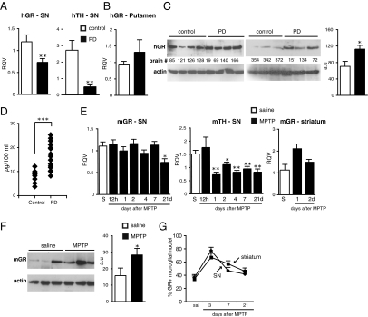
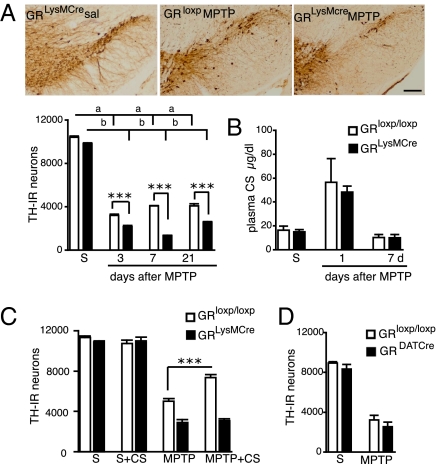
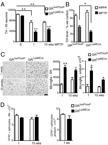
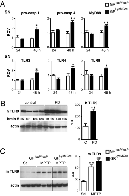
Among the pathogenic processes contributing to dopaminergic neuron (DN) death in Parkinson disease (PD), evidence points to non-cell-autonomous mechanisms, particularly chronic inflammation mounted by activated microglia. Yet little is known about endogenous regulatory processes that determine microglial actions in pathological states. We examined the role of glucocorticoid receptors (GRs), activated by glucocorticoids released in response to stress and known to regulate inflammation, in DN survival. Overall GR level was decreased in substantia nigra of PD patients and 1-methyl-4-phenyl-1,2,3,6-tetrahydropyridine (MPTP)-intoxicated mice. GR changes, specifically in the microglia after MPTP treatment, revealed a rapid augmentation in the number of microglia displaying nuclear localization of GR. Mice with selective inactivation of the GR gene in macrophages/microglia (GR(LysMCre)) but not in DNs (GR(DATCre)) showed increased loss of DNs after MPTP intoxication. This DN loss in GR(LysMCre) mice was not prevented by corticosterone treatment, in contrast to the protection observed in control littermates. Moreover, absence of microglial GRs augmented microglial reactivity and led to their persistent activation. Analysis of inflammatory genes revealed an up-regulation of Toll-like receptors (TLRs) by MPTP treatment, particularly TLR9, the level of which was high in postmortem parkinsonian brains. The regulatory control of GR was reflected by higher expression of proinflammatory genes (e.g., TNF-α) with a concomitant decrease in anti-inflammatory genes (e.g., IL-1R2) in GR(LysMCre) mice. Indeed, in GR(LysMCre) mice, alterations in phosphorylated NF-κB levels indicated its protracted activation. Together, our data indicate that GR is important in curtailing microglial reactivity, and its deregulation in PD could lead to sustained inflammation-mediated DN injury.


Conflict of interest statement

The authors declare no conflict of interest.

Figures

Fig. 1.
Fig. 1.
GR expression in SN and striatum in PD and in MPTP-treated mice. RT-qPCR of human (h) GR and TH mRNA from SN (A) and putamen (B) of control and PD (n = 5–6). (C) Western blot analysis of human striatal GR protein of two groups of PD and control subjects. The results were quantified with actin as loading control, a.u., arbitrary units. (D) Plasma cortisol levels of control subjects (n = 9) and PD patients (n = 20). (E) Time course of mouse (m) GR and TH mRNA levels in SN after saline (S) or acute MPTP treatment. GR mRNA levels in striatum after saline (S) or 1 or 2 d after MPTP treatment. (F) A representative experiment of Western blot analysis of striatal GR protein levels in mice injected with saline (S) or 7 d after MPTP. (G) Percentage of microglia showing nuclear GR localization quantified from confocal images of GR, Iba-1, and DAPI immunofluorescence in SN and striatum of saline control and MPTP-intoxicated mice killed at indicated days. (n = 3–5 for saline group, and n = 4–5 for each MPTP group.) *P < 0.05, **P ≤ 0.01, ***P < 0.001 (human control subjects vs. PD; saline vs. MPTP-injected mice, Mann-Whitney test).
Fig. 2.
Fig. 2.
Microglial GR protects DNs after acute MPTP intoxication. (A) TH immunohistochemistry in SN of GRLysMCre and GRloxP/loxP mice 7 d after either saline (sal) or MPTP treatment. (Bar = 100 μm.) Quantification of the number of TH-IR neurons in SN shows a significant reduction in GRLysMCre mutants compared with GRloxP/loxP controls after MPTP injections. Two-way ANOVA analysis followed by post hoc Bonferroni/Dunn test showed statistical significance for genotype on MPTP treatment at all time points and significance of P = 0.01 with F = 9.3, degrees of freedom = 1 at 7 d for genotype × treatment. a, b = ***P < 0.001 saline vs. MPTP for GRloxP/loxP and GRLysMCre mice. ***P < 0.001, **P < 0.01 MPTP GRloxP/loxP vs. GRLysMCre MPTP mice. (B) Basal CS levels in saline-injected (S) and MPTP-treated control and mutant mice at times indicated. (C) GRloxP/loxP and GR LysMCre were treated with β-cyclodextrin or CS + β-cyclodextrin for 7 d after saline or acute MPTP intoxication. The results show number of TH-IR neurons in SN. ***P < 0.001. (D) TH-IR cells in SN in control GRloxP/loxP and GR DATCre mice after saline (S) or MPTP. (n = 5–7 mice per group and per time point.)
Fig. 3.
Fig. 3.
Absence of GR increases loss of TH-IR neurons in SN and chronically activates microglia in GRLysMCre mutant mice after subchronic MPTP treatment. (A) Quantification of TH-IR neurons in SN of saline (S) or subchronically MPTP-intoxicated control GRloxP/loxP and GRLysMCre mutant mice, 1 wk or 10 wk after last injection. Two-way ANOVA followed by post hoc Bonferroni/Dunn test showed genotype × MPTP treatment effect: P = 0.02, F = 0.47, degrees of freedom = 2 (n = 4–5). (B) DA levels 1 wk after MPTP or saline injections. The results are calculated as percentage change from values obtained in corresponding saline-injected mice. (C) Immunohistochemistry with anti–Iba-1 antibody in SN 1 and 10 wk after MPTP treatment shows strong microglial activation in the GR LysMCre mutants. (Bar = 50 μm.) The hypertrophied Iba1+ cells were quantified both in SN and striatum; the activation persists in GRLysMCre mutants at 10 wk. (D) Quantification of GFAP+ astrocytes in the SN and striatum show no difference between GRloxP/loxP control and GRLysMCre mutants. Note that GFAP+ cells are absent in striatum in controls and mutants 10 wk after MPTP treatment. *P < 0.05 (n = 5).
Fig. 4.
Fig. 4.
GR regulation of upstream activators of innate immune response and relevance to PD. (A) qPCR results show up-regulation in the mRNA levels in SN of proinflammatory caspases and TLRs in GRLysMCre mutant mice compared with controls after MPTP. *P < 0.05, **P < 0.01, MPTP-treated GRloxP/loxP vs. GRLysMCre mice (n = 4). (B) TLR9 protein levels in striatum of human control subjects (C) and parkinsonian patients (PD). **P < 0.01, control subjects vs. PD (n = 5). (C) A representative Western blot experiment showing striatal protein levels of TLR9 in GRloxP/loxP control and GRLysMCre mutant mice 7 d after either saline (sal) or acute MPTP treatment. The signals were quantified in relation to actin. **P < 0.01, saline vs. MPTP treatment (n = 5).

References

    1. Dauer W, Przedborski S. Parkinson's disease: Mechanisms and models. Neuron. 2003;39:889–909. - PubMed
    1. McGeer PL, Itagaki S, Akiyama H, McGeer EG. Rate of cell death in parkinsonism indicates active neuropathological process. Ann Neurol. 1988;24:574–576. - PubMed
    1. Kurkowska-Jastrzebska I, Wrońska A, Kohutnicka M, Członkowski A, Członkowska A. The inflammatory reaction following 1-methyl-4-phenyl-1,2,3, \6-tetrahydropyridine intoxication in mouse. Exp Neurol. 1999;156:50–61. - PubMed
    1. Benner EJ, et al. Nitrated α-synuclein immunity accelerates degeneration of nigral dopaminergic neurons. PLoS ONE. 2008;3:e1376. - PMC - PubMed
    1. Ouchi Y, et al. Microglial activation and dopamine terminal loss in early Parkinson's disease. Ann Neurol. 2005;57:168–175. - PubMed

Publication types

MeSH terms

Substances

LinkOut - more resources