Review
. 2011 Oct;17(5):524-38.
doi: 10.1177/1073858410392239. Epub 2011 Apr 5.

The mirror neuron system: a fresh view

Affiliations
Review

The mirror neuron system: a fresh view

Antonino Casile et al. Neuroscientist. 2011 Oct.

Abstract

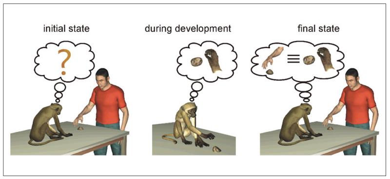
Mirror neurons are a class of visuomotor neurons in the monkey premotor and parietal cortices that discharge during the execution and observation of goal-directed motor acts. They are deemed to be at the basis of primates' social abilities. In this review, the authors provide a fresh view about two still open questions about mirror neurons. The first question is their possible functional role. By reviewing recent neurophysiological data, the authors suggest that mirror neurons might represent a flexible system that encodes observed actions in terms of several behaviorally relevant features. The second question concerns the possible developmental mechanisms responsible for their initial emergence. To provide a possible answer to question, the authors review two different aspects of sensorimotor development: facial and hand movements, respectively. The authors suggest that possibly two different "mirror" systems might underlie the development of action understanding and imitative abilities in the two cases. More specifically, a possibly prewired system already present at birth but shaped by the social environment might underlie the early development of facial imitative abilities. On the contrary, an experience-dependent system might subserve perception-action couplings in the case of hand movements. The development of this latter system might be critically dependent on the observation of own movements.


Figures

Figure 1
Figure 1
Areas in the monkey brain involved in action perception. The figure shows a lateral view of the left hemisphere of a macaque brain. The three highlighted areas contain neurons that discharge during observation of bodily movements. More specifically, neurons in the superior temporal sulcus (STS) exhibit purely visual responses and do not respond during the execution of movements. Neurons responding both during the observation and execution of goal-directed movements (i.e., mirror neurons) were found in areas F5c and PFG.
Figure 2
Figure 2
Response properties of a mirror neuron. Each row in the left column schematically represents the experimental condition. The panels in the right column show the corresponding responses of the same neuron in the form of a raster plot (upper part) and peri-stimulus spike density (bottom part). (top row, right panel) Response of a neuron during active goal-directed motor acts of the monkey (e.g., grasping small objects of different shapes). (bottom row, right panel) Response of the same neuron during the observation of the same goal-directed motor acts performed by the experimenter. In the two raster plots, each vertical bar signifies the occurrence of an action potential, and different lines refer to different trials. In both figures, time t = 0 represents the moment of contact between the monkey’s (top panel) or experimenter’s (bottom panel) hand with the goal object.
Figure 3
Figure 3
Mirror neurons encoding the intention of the actor. The four rows show the motor (first two rows) and the visual (third and fourth rows) responses of the same neuron under different experimental conditions, which are exemplified in the left column. The first two rows show the motor responses of a neuron when the monkey grasped a piece of food to eat it (first row) or to place it into a container (second row). The third and fourth rows show the discharges of the same neuron when the monkey was observing the experimenter grasping a piece of food to bring it to the mouth (third row) or to place it into a container (fourth row). During both action execution and observation, the unit discharged selectively only when the piece of food was grasped to eat it. It did not respond when the same object was grasped to place it into a container.
Figure 4
Figure 4
Mirror neurons encoding the spatial position of an observed action. The three rows show the discharges of two neurons (neuron 1 and neuron 2) during the execution of goal-directed hand movements (top row) and during the observation of actions executed in the extra- (middle row) and peri-personal (bottom row) space of the monkey, respectively. The yellow circle represents the spatial extent of the peri-personal space of the monkey. Both neuron 1 and neuron 2 responded during the execution of goal-directed movements (top row). However, they exhibited opposite behaviors during the observation of actions performed by the experimenter in front of the monkeys. Neuron 1 discharged more strongly during the observation of an action executed in the extra-personal space of the monkey. On the contrary, neuron 2 exhibited a significantly stronger discharge when the observed action was executed in the peri-personal space of the monkey.
Figure 5
Figure 5
Exemplification of the proposed mechanism for the initial emergence of mirror neurons. At the beginning of development, there is in the infant (human or monkey) no clear mapping between the observed motor act and its internal motor representation (left panel). During development, the repeated synchronous coactivation of motor and visual representations during observation of own movements leads to the emergence of neurons that exhibit a visuomotor coupling between a specific action and its visual representation from an egocentric point of view (middle panel). These visual representations are then generalized to the actions of others, which can be seen under a multiplicity of points of view (right panel).

References

    1. Abravanel E, Sigafoos AD. Exploring the presence of mitation during early infancy. Child Dev. 1984;55(2):381–92. Retrieved from: http://www.ncbi.nlm.nih.gov/pubmed/6723442. - PubMed
    1. Aggarwal JK, Cai Q. Human motion analysis: a review. Comput Vision Image Understanding. 1999;73:428–40.
    1. Anisfeld M. Neonatal imitation. Dev Rev. 1991;11:60–97.
    1. Anisfeld M. Only tongue protrusion modeling is matched by neonates. Dev Rev 16. 1996;2:149–61.
    1. Anisfeld M, Masters JC, Jacobson SW, Kagan J, Meltzoff AN, Moore MK. Interpreting “imitative” responses in early infancy. Science. 1979;205:214–9. Retrieved from: http://images2.wikia.nocookie.net/imitation/images/9/9f/Anisfeld_et_al_1.... - PubMed

Publication types

LinkOut - more resources