. 2011 Jun;188(2):349-58.
doi: 10.1534/genetics.111.128827. Epub 2011 Apr 5.

A forward genetic screen identifies mutants deficient for mitochondrial complex I assembly in Chlamydomonas reinhardtii

Affiliations

A forward genetic screen identifies mutants deficient for mitochondrial complex I assembly in Chlamydomonas reinhardtii

M Rosario Barbieri et al. Genetics. 2011 Jun.

Abstract

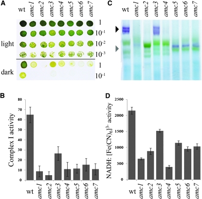
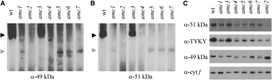
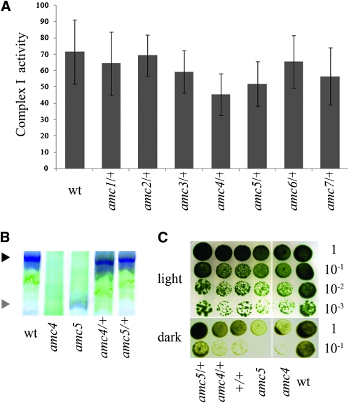
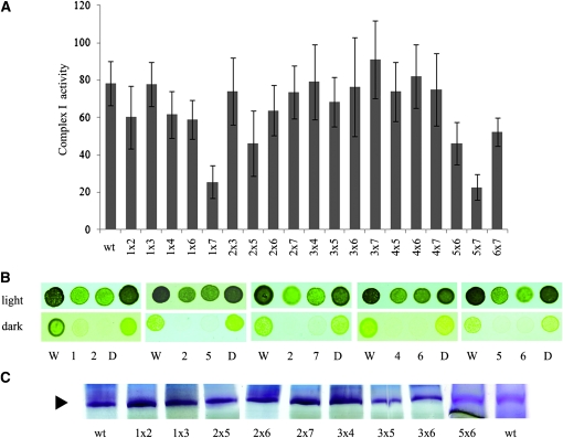
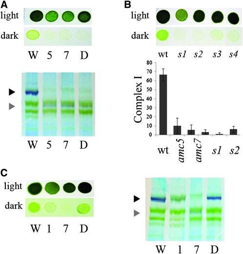
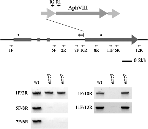
Mitochondrial complex I is the largest multimeric enzyme of the respiratory chain. The lack of a model system with facile genetics has limited the molecular dissection of complex I assembly. Using Chlamydomonas reinhardtii as an experimental system to screen for complex I defects, we isolated, via forward genetics, amc1-7 nuclear mutants (for assembly of mitochondrial complex I) displaying reduced or no complex I activity. Blue native (BN)-PAGE and immunoblot analyses revealed that amc3 and amc4 accumulate reduced levels of the complex I holoenzyme (950 kDa) while all other amc mutants fail to accumulate a mature complex. In amc1, -2, -5-7, the detection of a 700 kDa subcomplex retaining NADH dehydrogenase activity indicates an arrest in the assembly process. Genetic analyses established that amc5 and amc7 are alleles of the same locus while amc1-4 and amc6 define distinct complementation groups. The locus defined by the amc5 and amc7 alleles corresponds to the NUOB10 gene, encoding PDSW, a subunit of the membrane arm of complex I. This is the first report of a forward genetic screen yielding the isolation of complex I mutants. This work illustrates the potential of using Chlamydomonas as a genetically tractable organism to decipher complex I manufacture.


Figures

F<sc>igure</sc> 1.—
Figure 1.—
Six novel sid mutants display a defect in complex I activity. (A) Tenfold dilution series of amc17 and wild-type (wt) cells were plated on acetate-containing medium and incubated 3 days in the light or 5 days in the dark. (B) Complex I-specific activity (nmol NADH reduced/min/mg of protein) is indicated as averages of 10 independent measurements. (C) BN-PAGE and in-gel NBT staining of NADH dehydrogenase activity. The black and gray arrowheads indicate the position of the mature 950-kDa complex I and the 700-kDa subcomplex, respectively. The purple bands indicate staining for NADH oxidase activity in complex I and subcomplexes. Note that complex I is often resolved into two or more purple bands of high molecular weight. The identity of such bands is unclear but they do not correspond to the supercomplex I + III2 (Cardol et al. 2008). The green bands correspond to photosystems I and II and their respective light harvesting complexes of the chloroplast (Rexroth et al. 2003; Cardol et al. 2006). (D) NADH:ferricyanide oxidoreductase activity (nmol K3[Fe (CN)6] reduced/min/mg of protein). The results represent the average of three independent determinations. (B and D) Error bars indicate standard error (SE).
F<sc>igure</sc> 2.—
Figure 2.—
The amc mutants are deficient in complex I assembly. (A and B) Immunoblot analyses of membrane fractions, separated by BN-PAGE, using anti-49 kDa (A) and anti-51 kDa (B). A total of 150 μg of proteins was loaded per lane. The figures show the most representative blots of three independent experiments. The solid and shaded arrowheads indicate the position of mature complex I and the 700-kDa subcomplex, respectively. Equal loading was tested by Coomassie blue staining (not shown) and the presence of a nonspecific band revealed by the anti–49-kDa antiserum (Figure S2B for an example). (C) Membrane fractions were separated by SDS–PAGE and immunoblot analyses were performed using polyclonal antibodies against subunit-specific peptides. The subunits are 51 kDa, TYKY, and 49 kDa. A total of 10 μg of total protein per lane was loaded. Antibody specificity was evaluated by peptide competition assays (not shown). Only the top band is specific for the 49-kDa subunit. Plastid cytochrome f was used as a loading control. The results are representative from at least three independent membrane extractions.
F<sc>igure</sc> 3.—
Figure 3.—
Effects of the amc mutations on other respiratory complexes. (A) Complex II activity (succinate:2,6-dichlorophenolindophenol (DCIP) oxidoreductase). Three independent determinations were averaged and activities are expressed as nmol DCIP reduced/min/mg of protein. (B) Complex III activity (decylubiquinol:cytochrome c oxidoreductase). Averages from three independent measurements are shown in nmol of cytochrome c reduced/min/mg of protein. (C) Complexes II + III combined activities (succinate:cytochrome c oxidoreductase) of 10 independent determinations (nmol of cytochrome c reduced/min/mg of protein). dum11 is a complex III mutant (Dorthu et al. 1992). (D) Complex IV activity (cytochrome c oxidase). dum18 is a complex IV mutant (Remacle et al. 2001b). Average activities of 10 independent determinations are expressed in nmol cytochrome c oxidized/min/mg of protein. Error bars indicate SE.
F<sc>igure</sc> 4.—
Figure 4.—
The amc mutations are recessive. Heterozygous (amc/+) and homozygous (+/+) diploid strains were generated by crosses. (A) Complex I activities (nmol NADH oxidized/min/mg of protein). Columns represent the average of 10 independent determinations, with error bars representing SE. The +/+ diploid activity is not shown here and was 92 ± 8 nmol NADH oxidized/min/mg of protein. (B) BN-PAGE and in-gel NBT staining of NADH dehydrogenase activity. The black and gray arrowheads indicate the position of mature complex I and 700-kDa subcomplex, respectively. (C). Light/dark growth comparison of wild type (wt), amc4 and amc5 mutants, and their respective diploids (+/+, amc4/+, and amc5/+). Dilution series were plated on acetate containing medium and incubated 6 days in the light or 12 days in the dark. For B and C, only representative amc/+ diploids are shown. All amc/+ diploids exhibit restoration of the growth in the dark and complex I assembly phenotypes (not shown).
F<sc>igure</sc> 5.—
Figure 5.—
Complementation experiments define alleles of AMC loci. (A) Complex I activity of the wild type (wt) and the 19 diploids strains (amc × amc). The average of 10 independent replicas is indicated. The error bars represent standard error. The activity is expressed in nmol NADH oxidized/min/mg of protein. (B) Light/dark growth comparison of wild type (W), amc mutants (1, 2, and 47) and respective heterozygous diploids (D). Cells were plated on acetate-containing medium and incubated 3 days in the light or 5 days in the dark. (C) BN-PAGE and in-gel NBT staining of NADH dehydrogenase activity. The black arrowhead indicates the position of mature complex I. For B and C, only representative amc × amc diploids are shown. All diploids exhibiting restoration of the growth in the dark phenotype also showed restoration of complex I assembly as assessed by NBT staining (not shown). Only the top part of the BN gel is shown in C.
F<sc>igure</sc> 6.—
Figure 6.—
amc5 and amc7 define alleles of the same AMC locus. (A, top) Light/dark growth comparison of wild type (W), amc5 and amc7, and the amc5/amc7 diploid (D). (A, bottom) Detection of complex I and the 700-kDa subcomplex via NADH dehydrogenase activity on membrane fractions separated by BN-PAGE. (B, top) Light vs. dark growth of wild type (wt) and representative spores (s1s4) from the amc5 × amc7 cross. Note that the strains used in our study are not isogenic and it is possible that other genetic factors segregate and modulate the slow growth phenotype (compare s1 and s2 to s3 and s4). (B, bottom) Complex I activities (nmol NADH oxidized/min/mg of protein) of wild type (wt), amc5, amc7, and two sid spores (s1 and s2) originating from the amc5 × amc7 cross. Columns represent the average of 10 independent determinations, with SE as the error bars. (C, left) Light vs. dark growth of wild type (W), amc1 and amc7 mutants, and the amc1/amc7 diploid (D). (C, right) Detection of complex I and the 700-kDa subcomplex via NADH dehydrogenase activity on membrane fractions separated by BN-PAGE. (A, B, and C) Cells were plated on acetate-containing medium and incubated 3 days in the light or 5 days in the dark. (A and C) The black and gray arrowheads indicate the position of mature complex I and the 700-kDa subcomplex, respectively.
F<sc>igure</sc> 7.—
Figure 7.—
The amc5 and amc7 mutants carry molecular lesions in the NUOB10 gene encoding the noncore subunit PDSW. (Top) Schematic representation of the NUOB10 gene structure showing the four exons (rectangles) and three introns (thin lines). The asterisk denotes the position of the ATG codon while the “x” symbol indicates the position of the stop codon. The insertional cassette (AphVIII) is represented as an arrow (dark shading) flanked by the promoter and terminator regions (light shading). The positions of primers used to diagnose the molecular lesion are indicated by small arrows. The leftward arrow with a vertical line above intron 3 indicates the extent of the NUOB10 sequence obtained by TAIL-PCR (Figure S3). R1 and R2 indicate the positions of the AphVIII-specific primers used in the TAIL-PCR to recover the sequence flanking the cassette. A deletion of the promoter occurred upon integration of the cassette in the NUOB10 locus. (Bottom) Molecular lesions in NUOB10 were analyzed by PCR using NUOB10-specific primers. PCR products were stained by ethidium bromide and imaged using an imaging system (Kodak Image Station 2000R).

References

    1. Barrientos A., 2003. Yeast models of human mitochondrial diseases. IUBMB Life 55: 83–95 - PubMed
    1. Berthold P., Schmitt R., Mages W., 2002. An engineered Streptomyces hygroscopicus aph 7 gene mediates dominant resistance against hygromycin B in Chlamydomonas reinhardtii. Protist 153: 401–412 - PubMed
    1. Braun H.-P., Zabaleta E., 2007. Carbonic anhydrase subunits of the mitochondrial NADH dehydrogenase complex (complex I) in plants. Physiol. Plant. 129: 114–122
    1. Bych K., Kerscher S., Netz D. J., Pierik A. J., Zwicker K., et al. , 2008. The iron-sulphur protein Ind1 is required for effective complex I assembly. EMBO J. 27: 1736–1746 - PMC - PubMed
    1. Cardol P., Matagne R. F., Remacle C., 2002. Impact of mutations affecting ND mitochondria-encoded subunits on the activity and assembly of complex I in Chlamydomonas. Implication for the structural organization of the enzyme. J. Mol. Biol. 319: 1211–1221 - PubMed

Publication types

MeSH terms

Substances