. 2011 Apr;23(4):1449-67.
doi: 10.1105/tpc.110.082503. Epub 2011 Apr 5.

GUN4-porphyrin complexes bind the ChlH/GUN5 subunit of Mg-Chelatase and promote chlorophyll biosynthesis in Arabidopsis

Affiliations

GUN4-porphyrin complexes bind the ChlH/GUN5 subunit of Mg-Chelatase and promote chlorophyll biosynthesis in Arabidopsis

Neil D Adhikari et al. Plant Cell. 2011 Apr.

Abstract

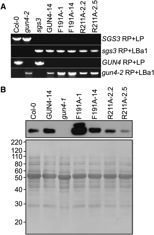
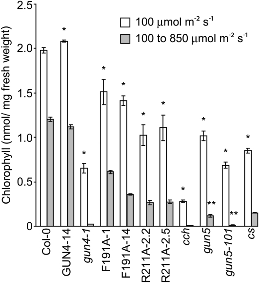
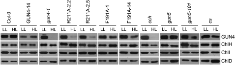
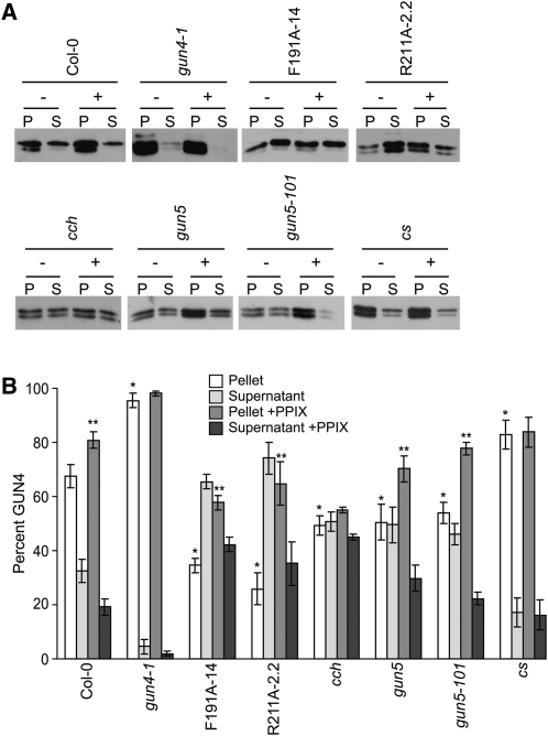
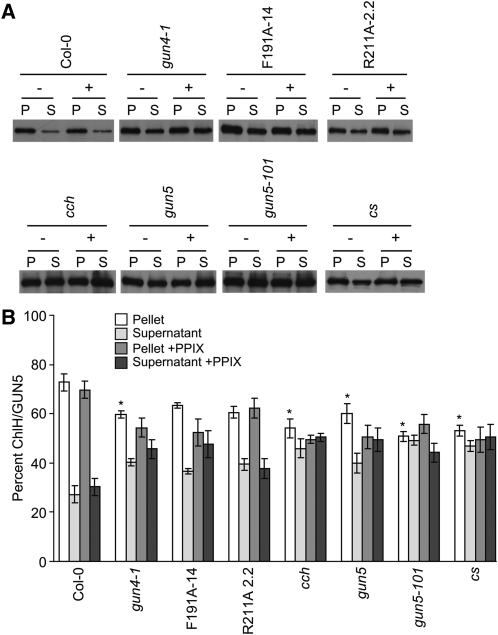
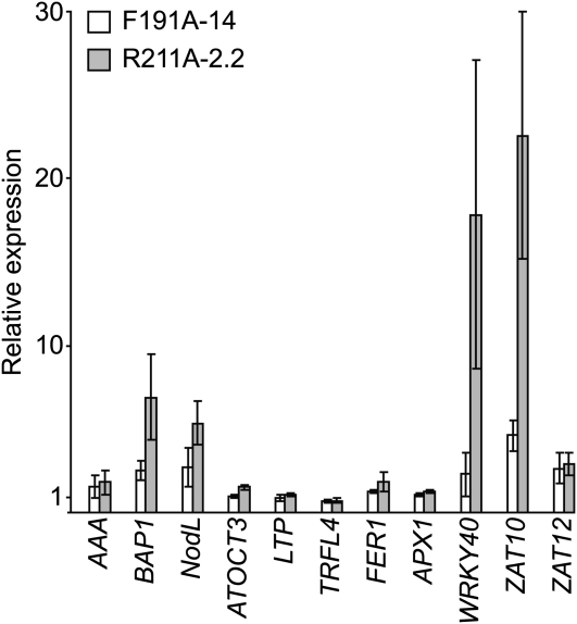
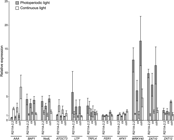
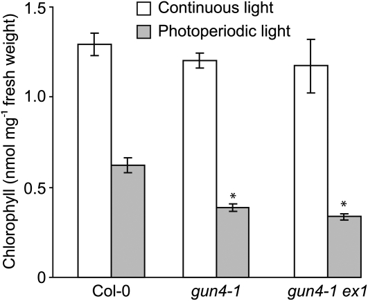
The GENOMES UNCOUPLED4 (GUN4) protein stimulates chlorophyll biosynthesis by activating Mg-chelatase, the enzyme that commits protoporphyrin IX to chlorophyll biosynthesis. This stimulation depends on GUN4 binding the ChlH subunit of Mg-chelatase and the porphyrin substrate and product of Mg-chelatase. After binding porphyrins, GUN4 associates more stably with chloroplast membranes and was proposed to promote interactions between ChlH and chloroplast membranes-the site of Mg-chelatase activity. GUN4 was also proposed to attenuate the production of reactive oxygen species (ROS) by binding and shielding light-exposed porphyrins from collisions with O₂. To test these proposals, we first engineered Arabidopsis thaliana plants that express only porphyrin binding-deficient forms of GUN4. Using these transgenic plants and particular mutants, we found that the porphyrin binding activity of GUN4 and Mg-chelatase contribute to the accumulation of chlorophyll, GUN4, and Mg-chelatase subunits. Also, we found that the porphyrin binding activity of GUN4 and Mg-chelatase affect the associations of GUN4 and ChlH with chloroplast membranes and have various effects on the expression of ROS-inducible genes. Based on our findings, we conclude that ChlH and GUN4 use distinct mechanisms to associate with chloroplast membranes and that mutant alleles of GUN4 and Mg-chelatase genes cause sensitivity to intense light by a mechanism that is potentially complex.


Figures

Figure 1.
Figure 1.
Analysis of Arabidopsis Plants That Were Stably Transformed with GUN4-Derived Transgenes. (A) Genotyping of the sgs3 and gun4-2 T-DNA insertion alleles in Arabidopsis plants that were stably transformed with GUN4-derived transgenes. Transgenic plants were homozygous for gun4-2, sgs3, the transgene indicated above lanes 4 to 8. Genomic DNA was extracted from the wild type (Col-0), the indicated mutant, or the indicated stably transformed line. Plants were screened for wild-type and T-DNA insertion alleles by PCR-based genotyping with oligonucleotides that can amplify PCR products from SGS3 (SGS3 RP + LP), GUN4 (GUN4 RP +LP), or particular T-DNA insertion alleles (sgs3 RP + LBa1 or gun4-2 RP + LBa1). PCR products were analyzed by electrophoresis in agarose gels followed by staining with ethidium bromide. (B) Analysis of GUN4 protein levels in Arabidopsis plants that are stably transformed with GUN4-derived transgenes. Transgenic plants were homozygous for gun4-2, sgs3, and the indicated transgene. Whole seedling extracts were prepared from the indicated mutant or the indicated stably transformed line grown in 100 μmol m−2 s−1 broad-spectrum white light. Aliquots of these whole seedling extracts that contained 10 μg of protein were analyzed by immunoblotting using anti-GUN4 antibodies (top panel) to detect the 22-kD band that corresponds to GUN4. After immunoblotting, the polyvinylidene fluoride membrane was stained with Coomassie blue (bottom panel). Mass standards are indicated at the left in kilodaltons.
Figure 2.
Figure 2.
Analysis of Chlorophyll Levels in gun4, chlH/gun5, and cs Mutants Grown under Various Fluence Rates. Wild type (Col-0) and the indicated mutants and transgenic lines were grown in continuous 100 μmol m−2 s−1 white light for 7 d (white bars) or for 3 d in continuous 100 μmol m−2 s−1 white light and then 4 d in continuous 850 μmol m−2 s−1 white light (gray bars). Chlorophyll was extracted from at least three biological replicates for each mutant or line in each condition. Error bars indicate se. The statistical significance of the difference between the chlorophyll levels of the wild type (Col-0) and a particular mutant or transgenic line was calculated with an unpaired t test. A single asterisk indicates a statistically significant difference in chlorophyll levels between Col-0 and a particular mutant or transgenic line (P = 0.0001 to 0.03). Using an unpaired t test, we calculated the statistical significance of the decrease in chlorophyll levels following the fluence rate shift from 100 μmol m−2 s−1 to 850 μmol m−2 s−1 white light between (1) gun5-101 and gun4-1 and (2) between gun5 and the following mutants and transgenic lines: cs, R211A-2.2, and R211A-2.5. A double asterisk indicates a statistically significant difference (P = 0.003 to 0.04).
Figure 3.
Figure 3.
Analysis of GUN4 and Mg-Chelatase Subunit Levels in 100 μmol m−2 s−1 and 850 μmol m−2 s−1 White Light. Seedlings were grown either in 100 μmol m−2 s−1 white light for 7 d (LL) or in 100 μmol m−2 s−1 for 3 d and then transferred 850 μmol m−2 s−1 white light for 4 d (HL) as indicated in Figure 2. Whole seedling extracts were prepared from the indicated mutant or the indicated stably transformed line. Aliquots of these whole seedling extracts that contained 10 μg of protein were analyzed by immunoblotting using the following antibodies: anti-GUN4, anti-ChlH, anti-ChlI, or anti-ChlD. When faint bands were observed in extracts from HL-treated seedlings, exposures were adjusted so that these faint bands were observable.
Figure 4.
Figure 4.
Distribution of GUN4 in Lysed and Fractionated Chloroplasts That Were Either Fed or Not Fed with PPIX. (A) Immunoblot analysis of lysed and fractionated chloroplasts using anti-GUN4 antibodies. Chloroplasts were purified from gun4-1, F191A-14, R211A-2.2, cch, gun5, gun5-101, and cs. Purified intact chloroplasts (200 μg) were either fed (+) or not fed (−) with 20 μM PPIX. Chloroplasts were then fractionated into soluble (S) and membrane-containing pellet (P) fractions of equal volume. Equal volumes were analyzed by SDS-PAGE and immunoblotting with anti-GUN4 antibodies. Representative immunoblots are shown. (B) Proportions of GUN4 in soluble and pellet fractions. The chemiluminescence that was emitted from the immunoreactive bands described in (A) was quantified. The percentage of GUN4 in the pellet (white bars) and supernatant (light-gray bars) fractions derived from chloroplasts that were not fed PPIX and the percentage of GUN4 in the pellet (medium-gray bars) and supernatant (dark-gray bars) fractions derived from chloroplasts that were fed PPIX are indicated for wild type (Col-0) and each mutant and transgenic line. Results from at least five independent experiments for Col-0 and four independent experiments for all mutants and transgenic lines are shown. Error bars indicate se. A single asterisk indicates a statistically significant difference in the levels of GUN4 in the membrane-containing pellet fractions between Col-0 and the gun4 mutants gun4-1, F191A-14, and R211A-2.2 (P < 0.0005) and between Col-0 and the chlH/gun5 mutants cch (P < 0.01), gun5, gun5-101, and cs (P < 0.04). A double asterisk indicates a statistically significant difference in the percent GUN4 in the membrane-containing pellet fractions derived from chloroplasts that were fed PPIX and not fed PPIX and were purified from Col-0 (P = 0.009), F191A-14 (P = 0.002), R211A-2.2 (P = 0.002), gun5 (P = 0.04), and gun5-101 (P = 0.007).
Figure 5.
Figure 5.
Distribution of ChlH/GUN5 in Lysed and Fractionated Chloroplasts That Were Either Fed or Not fed with PPIX. (A) Analysis of lysed and fractionated chloroplasts by immunoblotting with anti-ChlH/GUN5 antibodies. The same fractions described in Figure 4 were analyzed by immunoblotting with anti-ChlH/GUN5 antibodies. As described for Figure 4, 200 μg of purified intact chloroplasts were either fed (+) or not fed (−) with 20 μM PPIX. These chloroplasts were then fractionated into soluble (S) and membrane-containing pellet (P) fractions of equal volume. Equal volumes were analyzed by SDS-PAGE and immunoblotting. (B) Proportions of ChlH/GUN5 in soluble and membrane fractions. The percentage of ChlH/GUN5 in the pellet (white bars) and supernatant (light-gray bars) fractions derived from chloroplasts that were not fed PPIX and the percentage of ChlH/GUN5 in the pellet (medium-gray bars) and supernatant (dark-gray bars) fractions derived from chloroplasts that were fed PPIX are indicated for the wild type (Col-0) and each mutant and transgenic line. Results from seven independent experiments are shown for Col-0, five independent experiments are shown for cs, and four independent experiments are shown for all other mutants and transgenic lines. Error bars indicate se. The asterisk indicates a statistically significant difference in the levels of ChlH/GUN5 in the membrane-containing pellet fractions between Col-0 and gun4-1 (P = 0.02), between Col-0 and cch (P = 0.008), and between gun5 (P = 0.04), gun5-101, and cs (P < 0.002).
Figure 6.
Figure 6.
Analysis of ROS-Regulated Gene Expression in F191A and R211A Grown in 10 μmol m−2 s−1 White Light. The wild type (Col-0), F191A-14, and R211A-2.2 were grown for 7 d in 10 μmol m−2 s−1 white light. Levels of transcripts indicated were quantified by means of qRT-PCR. Expression in F191A-14 (white bars) and R211A-2.2 (gray bars) is reported relative to Col-0, which is assigned a value of 1. Four biological replicates were analyzed for each line. Error bars represent se.
Figure 7.
Figure 7.
Analysis of ROS-Inducible Gene Expression in Photoperiodic and Continuous Light. Seedlings were grown for 7 d in 12 h of 2 μmol m−2 s−1 white light followed by 12 h darkness (gray bars) or in continuous 2 μmol m−2 s−1 white light (white bars). All seedlings were harvested 1 h after dawn. The expression of ROS-inducible genes was quantified by means of qRT-PCR as described in Figure 6. Expression is reported relative to Col-0 in continuous light, which was assigned a value of 1. Four biological replicates were analyzed for each line in each condition. Error bars represent se.
Figure 8.
Figure 8.
Quantitative Analysis of Chlorophyll Levels in the Wild Type (Col-0), gun4-1, and gun4-1 ex1. Wild-type (Col-0), gun4-1, and gun4-1 ex1 seedlings were grown for 14 d in a photoperiod in which 12 h of 10 μmol m−2 s−1 white light was followed by 12 h of darkness (gray bars) or in continuous 10 μmol m−2 s−1 white light (white bars). Four biological replicates were collected for each line in each condition. The statistical significance of the difference between the levels of chlorophyll in the wild type (Col-0) and a particular mutant was calculated with an unpaired t test. Error bars indicate se. The asterisk indicates a statistically significant difference in the levels of chlorophyll between the wild type (Col-0) and gun4-1 (P = 0.002) and between the wild type and gun4-1 ex1 (P = 0006) grown in photoperiodic light.

References

    1. Aarti D., Tanaka R., Ito H., Tanaka A. (2007). High light inhibits chlorophyll biosynthesis at the level of 5-aminolevulinate synthesis during de-etiolation in cucumber (Cucumis sativus) cotyledons. Photochem. Photobiol. 83: 171–176 - PubMed
    1. Aarti D.P., Tanaka R., Tanaka A. (2006). Effects of oxidative stress on chlorophyll biosynthesis in cucumber (Cucumis sativus) cotyledons. Physiol. Plant. 128: 186–197
    1. Adhikari N.D., Orler R., Chory J., Froehlich J.E., Larkin R.M. (2009). Porphyrins promote the association of GENOMES UNCOUPLED 4 and a Mg-chelatase subunit with chloroplast membranes. J. Biol. Chem. 284: 24783–24796 - PMC - PubMed
    1. Alawady A.E., Grimm B. (2005). Tobacco Mg protoporphyrin IX methyltransferase is involved in inverse activation of Mg porphyrin and protoheme synthesis. Plant J. 41: 282–290 - PubMed
    1. Allen K.D., Duysen M.E., Staehelin L.A. (1988). Biogenesis of thylakoid membranes is controlled by light intensity in the conditional chlorophyll b-deficient CD3 mutant of wheat. J. Cell Biol. 107: 907–919 - PMC - PubMed

Publication types

MeSH terms