

Noninvasive In vivo assessment of renal tissue elasticity during graded renal ischemia using MR elastography
- PMID: 21467945
- PMCID: PMC3128234
- DOI: 10.1097/RLI.0b013e3182183a95
Noninvasive In vivo assessment of renal tissue elasticity during graded renal ischemia using MR elastography
Abstract
Objectives: : Magnetic resonance elastography (MRE) allows noninvasive assessment of tissue stiffness in vivo. Renal arterial stenosis (RAS), a narrowing of the renal artery, promotes irreversible tissue fibrosis that threatens kidney viability and may elevate tissue stiffness. However, kidney stiffness may also be affected by hemodynamic factors. This study tested the hypothesis that renal blood flow (RBF) is an important determinant of renal stiffness as measured by MRE.
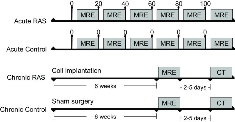
Material and methods: : In 6 anesthetized pigs MRE studies were performed to determine cortical and medullary elasticity during acute graded decreases in RBF (by 20%, 40%, 60%, 80%, and 100% of baseline) achieved by a vascular occluder. Three sham-operated swine served as time control. Additional pigs were studied with MRE 6 weeks after induction of chronic unilateral RAS (n = 6) or control (n = 3). Kidney fibrosis was subsequently evaluated histologically by trichrome staining.
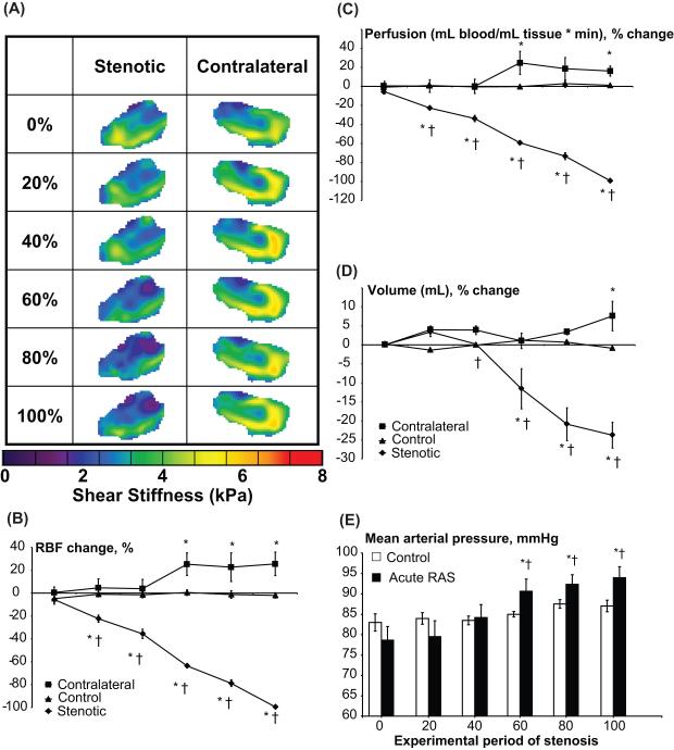
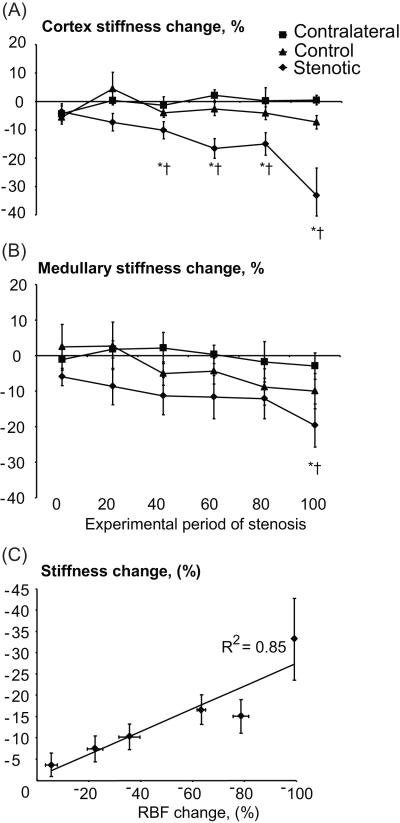
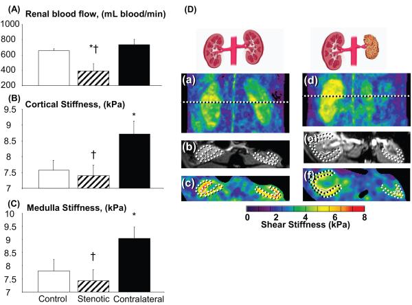
Results: : During acute RAS the stenotic cortex stiffness decreased (from 7.4 ± 0.3 to 4.8 ± 0.6 kPa, P = 0.02 vs. baseline) as RBF decreased. Furthermore, in pigs with chronic RAS (80% ± 5.4% stenosis) in which RBF was decreased by 60% ± 14% compared with controls, cortical stiffness was not significantly different from normal (7.4 ± 0.3 vs. 7.6 ± 0.3 kPa, P = 0.3), despite histologic evidence of renal tissue fibrosis.
Conclusion: : Hemodynamic variables modulate kidney stiffness measured by MRE and may mask the presence of fibrosis. These results suggest that kidney turgor should be considered during interpretation of elasticity assessments.
Figures
References
-
- Schlondorff DO. Overview of factors contributing to the pathophysiology of progressive renal disease. Kidney Int. 2008;74:860–6. - PubMed
-
- Rouviere O, Yin M, Dresner MA, et al. MR elastography of the liver: preliminary results. Radiology. 2006;240:440–8. - PubMed
-
- Chade AR, Zhu XY, Grande JP, et al. Simvastatin abates development of renal fibrosis in experimental renovascular disease. J Hypertens. 2008;26:1651–60. - PubMed
-
- Stephens SE, Rigden SP. Cystic fibrosis and renal disease. Paediatr Respir Rev. 2002;3:135–8. - PubMed
Publication types
MeSH terms
Grants and funding
- R01 HL077131/HL/NHLBI NIH HHS/United States
- C06 RR018898/RR/NCRR NIH HHS/United States
- R21 DK077013/DK/NIDDK NIH HHS/United States
- 018898/PHS HHS/United States
- R37 EB001981/EB/NIBIB NIH HHS/United States
- HL085307/HL/NHLBI NIH HHS/United States
- 001981/PHS HHS/United States
- R01 DK073608/DK/NIDDK NIH HHS/United States
- R01 EB001981/EB/NIBIB NIH HHS/United States
- DK73013/DK/NIDDK NIH HHS/United States
- EB001981/EB/NIBIB NIH HHS/United States
- HL77131/HL/NHLBI NIH HHS/United States
- R01 EB002640/EB/NIBIB NIH HHS/United States
- P01 HL085307/HL/NHLBI NIH HHS/United States
- DK73608/DK/NIDDK NIH HHS/United States
LinkOut - more resources
Full Text Sources
