. 2011 Oct;21(10):1452-69.
doi: 10.1038/cr.2011.60. Epub 2011 Apr 5.

GPR55 regulates cannabinoid 2 receptor-mediated responses in human neutrophils

Affiliations

GPR55 regulates cannabinoid 2 receptor-mediated responses in human neutrophils

Nariman A B Balenga et al. Cell Res. 2011 Oct.

Erratum in

  • Cell Res. 2011 Nov;21(11):1641

Abstract

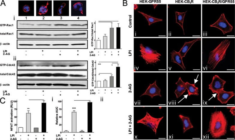
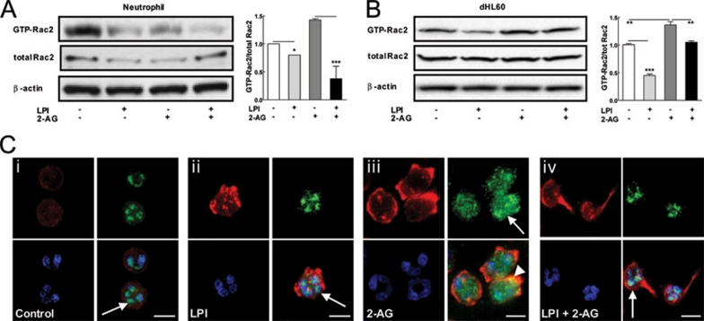
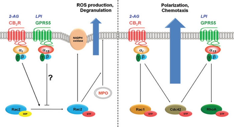
The directional migration of neutrophils towards inflammatory mediators, such as chemokines and cannabinoids, occurs via the activation of seven transmembrane G protein coupled receptors (7TM/GPCRs) and is a highly organized process. A crucial role for controlling neutrophil migration has been ascribed to the cannabinoid CB(2) receptor (CB(2)R), but additional modulatory sites distinct from CB(2)R have recently been suggested to impact CB(2)R-mediated effector functions in neutrophils. Here, we provide evidence that the recently de-orphanized 7TM/GPCR GPR55 potently modulates CB(2)R-mediated responses. We show that GPR55 is expressed in human blood neutrophils and its activation augments the migratory response towards the CB(2)R agonist 2-arachidonoylglycerol (2-AG), while inhibiting neutrophil degranulation and reactive oxygen species (ROS) production. Using HEK293 and HL60 cell lines, along with primary neutrophils, we show that GPR55 and CB(2)R interfere with each other's signaling pathways at the level of small GTPases, such as Rac2 and Cdc42. This ultimately leads to cellular polarization and efficient migration as well as abrogation of degranulation and ROS formation in neutrophils. Therefore, GPR55 limits the tissue-injuring inflammatory responses mediated by CB(2)R, while it synergizes with CB(2)R in recruiting neutrophils to sites of inflammation.


Figures

Figure 1
Figure 1
GPR55 and CB2R agonists induce the directional migration and polarization of neutrophils. (A) Human blood neutrophils were placed to the upper wells of a microBoyden chamber and (i) were allowed to migrate towards increasing concentrations of LPI (▪), AM251 (•) or 2-AG (♦) in the bottom wells for 1 h. Migrated cells in the bottom wells were counted by a flow cytometer. (ii) Neutrophils were pre-incubated with DMSO (0.05%), CBD (5 μM) or AM630 (5 μM) for 10 min at 37 °C and their migration towards LPI (3 μM) or 2-AG (1 μM) was assessed as in panel Ai. (iii) Chemotaxis of neutrophils towards DMSO (0.01%, white bar), LPI (3 μM, light gray bar), 2-AG (1 μM, dark gray bar) or LPI and 2-AG combined (3 μM and 1 μM, respectively, black bar) was assessed as in panel Ai. (iv) Chemotaxis of neutrophils was assessed as in panel iii, except that AM251 (3 μM) was used instead of LPI. (v) Neutrophils were pre-incubated with DMSO (0.05%), CBD (5 μM) and/or AM630 (5 μM) for 10 min at 37 °C and their migration towards DMSO (0.01%) and LPI (3 μM) + 2-AG (1 μM) was assessed as in panel Ai. Representatives of 3-6 independent experiments, performed in quadruplicates, are shown for all subpanels. Data are mean±SEM (*P< 0.05; **P< 0.01; ***P< 0.001). (B) Neutrophils were seeded on fibronectin-coated glass coverslips and treated with a gradient of 0.01% DMSO (control; i) or ligands for 5 min at 37 °C and stained with methanolic Texas-Red Phalloidin (red) and DAPI (blue). (ii) LPI treatment (3 μM) induced fuzzy protrusions (arrows), whereas (iii) 2-AG (1 μM) induced an elongation of the neutrophils (arrows). (iv) Extending head (arrow) and tail formation (dashed arrow) indicate a polarization of neutrophils in response to a mixture of LPI (3 μM) and 2-AG (1 μM). Cells were analyzed using a Zeiss LSM510 META Axioplan confocal microscope (original magnification: 100×). Scale bars: 10 μm. Representative images of three independent experiments are shown. (C) Neutrophils were seeded on fibronectin-coated glass coverslips and treated with LPI (3 μM) and 2-AG (1 μM) for 5 min at 37 °C and stained with Texas-Red Phalloidin (red) and DAPI (blue). (i) Cells displayed a clear directional and polarized structure when migrating towards a local source of LPI/2-AG (white dot). (ii) In contrast, no cytoskeleton remodeling occurred after a uniform addition of agonists to the medium. Cells were analyzed as in panel B (original magnification: 63×). Representative images of three independent experiments are shown. Scale bars: 10 μm.
Figure 2
Figure 2
GPR55 is highly expressed in human blood neutrophils and neutrophil-like HL60 cells. (A) GPR55 and CB2R mRNA expression in freshly isolated human blood neutrophils was assessed by (i) quantitative real-time PCR and (ii) RT-PCR. PCR products were analyzed on a 3% agarose gel. (iii) Relative expression of GPR55 mRNA in undifferentiated (uHL60) and HL60 cells differentiated with 1.75% DMSO for 4 days (dHL60) was measured by real-time PCR. Representatives of three independent experiments are shown for all subpanels. Data are mean±SEM (**P< 0.01). (B) (i) GPR55 and CB2R protein expression in neutrophils, uHL60 and dHL60 cells was assessed by western blotting using rat anti-GPR55 and rabbit anti-CB2R antibodies. Lysates were probed for β-actin as a loading control. A representative blot of three independent experiments is shown. (ii) Western blotting of GPR55 and CB2R in lysates from HEK293, HEK-GPR55, HEK-CB2R and HEK-CB2R/GPR55 cells was performed as in panel Bi. A representative blot of three independent experiments is shown. (C) GPR55 expression in human blood neutrophils, uHL60 and dHL60 cells was confirmed with the rat anti-GPR55 antibody (1:250) and an Alexa Fluor-594 goat anti-rat secondary antibody (1:250). The dHL60 cells show multilobular nuclei (arrows). Cells were analyzed using a Zeiss LSM510 META Axioplan confocal microscope (original magnification: 100×). Scale bars: 10 μm. Representative images of 2-3 independent experiments are shown.
Figure 3
Figure 3
GPR55 and CB2R mediate chemotaxis and cytoskeletal remodeling via coupling to Gα13/RhoA and Gαi proteins. Neutrophils were pre-incubated with cell-permeable C3 toxin (C3, 3 μg/ml) or pertussis toxin (PTX, 3 μg/ml) for 2 h at 37 °C in PBG buffer. (A) Cells were allowed to migrate towards (i) LPI (3 μM), (ii) 2-AG (1 μM) or (iii) a combination of LPI (3 μM) and 2-AG (1 μM) for 1 h. Migrated cells in the bottom wells were counted by a flow cytometer. The chemotactic index was calculated as number of cells migrated towards agonists divided by the number of cells migrated towards vehicle (DMSO 0.01%). Representatives of two independent experiments, performed in quadruplicates, are shown. Data are mean±SEM (*P< 0.05; **P< 0.01). n.s.: not significant. (B) Cells were seeded on fibronectin-coated glass coverslips and treated with 0.01% DMSO (Control), LPI (3 μM), 2-AG (1 μM) or a mixture of LPI (3 μM) and 2-AG (1 μM) for 5 min at 37 °C and stained with Texas-Red Phalloidin (red) and DAPI (blue). Cells were analyzed using an OLYMPUS fluorescence microscope equipped with a Hamamatsu ORCA CCD camera (original magnification: 60×). Scale bars: 10 μm. Representative images from 2 independent experiments are shown.
Figure 4
Figure 4
Cytoskeletal rearrangement of (A) neutrophils and (B) HEK293 cells requires the concomitant activation of GPR55 and CB2R. (A) Neutrophils were stimulated with LPI (1 μM), 2-AG (1 μM) and LPI (1 μM) + 2-AG (1 μM) for 1 min at 37 °C. Active GTP-bound Rac1 and Cdc42 GTPases were extracted from the lysates with PAK domain-gluthatione agarose beads. GTP-bound and total GTPase levels were visualized by western blotting using mouse anti-Rac1 (i) and rabbit anti-Cdc42 (ii) antibodies. The β-actin served as a loading control. The ratio of GTP-bound vs total GTPase levels was assessed with ImageJ software (graphs). Representative blots from 3-4 independent experiments are shown. Data are mean±SEM. (*P< 0.05; ***P< 0.001). (B) HEK-GPR55, HEK-CB2R and HEK-CB2R/GPR55 cells were seeded on 1% PDL-coated glass coverslips. Serum-starved cells were incubated with agonists (1 μM) for 10 min in a serum-free medium. The fixed cells were stained for F-actin by methanolic Texas-Red Phalloidin (red) and with DAPI (blue). Cells were analyzed using an OLYMPUS fluorescence microscope equipped with a Hamamatsu ORCA CCD camera (original magnification: 60×). Scale bars: 20 μm. Representative images from 3-4 experiments are shown. (C) (i) HEK-CB2R/GPR55 cells were transfected with 200 ng of NFAT-luciferase reporter plasmid and 24 h later cells were stimulated with agonists (1 μM) for 3 h in a serum-free medium. The luciferase activity was visualized using a steadylite plus kit (PerkinElmer). Luminescence (relative light units (RLU)) was measured in a TopCounter (Top Count NXT; Packard) for 5 s. Data are mean±SEM from three independent experiments performed in quadruplicate (**P< 0.01) (ii) HEK-CB2R/GPR55 cells were challenged with ligands (1 μM) and the resulting picometer shifts of reflected light wavelength against the time (s) were monitored. Transformation of optical signatures were made by using the area under the curve (AUC) values between the 1 200 and 3 600 s time points. Data were normalized and expressed as percent of maximum activation induced by LPI + 2-AG. Data are mean ±SEM from three independent experiments performed in quadruplicate (***P< 0.001).
Figure 5
Figure 5
GPR55 activation inhibits (A) CB2R-mediated respiratory burst and (B) C5a-induced degranulation in neutrophils. (A) ROS production in neutrophils was measured by flow cytometry. (i) Cells were loaded with 1 μM 2′,7′-DCF-DA and then incubated with DMSO (0.1%), LPI (300 nM), 2-AG (10 μM) or a combination of LPI and 2-AG for 20 min at 37 °C. ROS production was measured as a change in fluorescence in the FL1 channel. (ii) ROS production in neutrophils was measured as in panel Ai except that AM251 (300 nM) was used instead of LPI. (iii) Neutrophils were incubated with C5a (5 nM) or 2-AG (10 μM) and treated with buffer (control) or LPI (100 nM or 300 nM) for 20 min. ROS production was assessed as in panel Ai. (iv) Serum-starved dHL60 cells were loaded with 5 μM 2′,7′-DCF-DA for 10 min at 37 °C and then incubated with 2-AG (10 μM) in combination with assay buffer (control) or LPI (1 or 10 μM). LPI (10 μM) used in combination with DMSO (0.1%) did not induce changes in ROS levels. ROS production was recorded in a Flex-Station II device (Ex. 485nm, Em. 535 nm) 20 min after ligand addition. Representatives of 3-4 independent experiments, performed in quadruplicates are shown for all subpanels. Data are mean± SEM (*P< 0.05; **P< 0.01; ***P< 0.001). (B) (i) Neutrophils were incubated with LPI (300 nM) or assay buffer (control) for 1 h at 37 °C. MPO release was induced by increasing concentrations of C5a for 30 min and measured as the change in absorbance at 630 nm in a colorimetric assay. (ii) Neutrophils were incubated with increasing concentrations of LPI for 1 h at 37 °C. MPO release was induced with C5a (300 nM) for 30 min and assessed as in panel Bi. Data are mean±SEM of three independent experiments performed in triplicates (*P< 0.05; **P< 0.01; ***P< 0.001). The MPO release induced by 300 nM C5a was set to 100%.
Figure 6
Figure 6
GPR55 activation suppresses the CB2R-mediated activation and translocation of Rac2. (A) Neutrophils were stimulated with agonists (1 μM) for 1 min at 37 °C. The active GTP-bound Rac2 was extracted from the lysates with PAK domain-gluthatione agarose beads. GTP-bound and total GTPase levels were visualized by western blotting using a rabbit anti-Rac2 antibody, β-actin served as a loading control. The ratio of GTP-bound vs total GTPase levels was assessed with ImageJ software (graph). Data are mean±SEM of three independent experiments. (*P< 0.05; ***P< 0.001). (B) Serum-starved dHL60 cells were stimulated with agonists (1 μM) for 1 min at 37 °C. The extraction of active GTP-bound Rac2 was performed as in panel (A). Data are mean±SEM of three independent experiments (**P< 0.01; ***P< 0.001). (C) Neutrophils were seeded on fibronectin-coated glass coverslips and treated with 0.01% DMSO (control; i) or ligands for 5 min at 37 °C. Fixed cells were incubated with rabbit anti-Rac2 antibody and stained with Alexa Fluor-488 goat anti-rabbit antibody (green), Texas-Red Phalloidin (red), and DAPI (blue). Control (i) and LPI (3 μM, ii) treated cells show a nuclear/perinuclear localization of Rac2 (arrows). Upon 2-AG stimulation (1 μM, iii), Rac2 distributed evenly in the cytosol (arrow) and partially colocalized with actin at the plasma membrane (yellow, arrowhead). (iv) Treatment with a combination of LPI (3 μM) and 2-AG (1 μM) showed a nuclear localization of Rac2 in polarized neutrophils. Cells were analyzed using an OLYMPUS fluorescence microscope equipped with a Hamamatsu ORCA CCD camera (original magnification: 60×). Scale bars: 10 μm. Representative images from 2-3 experiments are shown.
Figure 7
Figure 7
Crosstalk between GPR55 and CB2R and its consequent biological responses in human blood neutrophils. (right panel) Stimulation of CB2R and GPR55 by 2-AG and LPI, respectively, leads to a coordinated activation of RhoA, Rac1 and Cdc42 small GTPases. This leads to a distinct remodeling of cytoskeleton (polarization) compared to sole activation of each receptor and facilitates neutrophils migration towards the gradient of agonists. (left panel) The bacterial killing mechanisms, which are provoked by C5a or 2-AG, are mediated via the activation of Rac2 small GTPases. Active Rac2 will translocate and incorporate to the NADPH oxidase core complex in the phagocytic cup and catalyzes the formation of reactive oxygen species (ROS). On the other hand, it facilitates degranulation of neutrophils via translocation of azurophilic granules, containing myeloperoxidase (MPO). Stimulation of GPR55 by LPI, via a yet unknown mechanism, inhibits activation of Rac2, thereby limiting the ROS production and degranulation.

Similar articles

Cited by

References

    1. Chakravarti A, Allaeys I, Poubelle PE. Neutrophils and immunity: is it innate or acquired. Med Sci (Paris) 2007;23:862–867. - PubMed
    1. Nathan C. Neutrophils and immunity: challenges and opportunities. Nat Rev Immunol. 2006;6:173–182. - PubMed
    1. Witko-Sarsat V, Rieu P, scamps-Latscha B, Lesavre P, Halbwachs-Mecarelli L. Neutrophils: molecules, functions and pathophysiological aspects. Lab Invest. 2000;80:617–653. - PubMed
    1. Luster AD, Alon R, von Andrian UH. Immune cell migration in inflammation: present and future therapeutic targets. Nat Immunol. 2005;6:1182–1190. - PubMed
    1. Monk PN, Scola AM, Madala P, Fairlie DP. Function, structure and therapeutic potential of complement C5a receptors. Br J Pharmacol. 2007;152:429–448. - PMC - PubMed

Publication types

MeSH terms

LinkOut - more resources