. 2011 Aug;19(8):1558-68.
doi: 10.1038/mt.2011.29. Epub 2011 Apr 5.

Optimizing DC vaccination by combination with oncolytic adenovirus coexpressing IL-12 and GM-CSF

Affiliations

Optimizing DC vaccination by combination with oncolytic adenovirus coexpressing IL-12 and GM-CSF

Song-Nan Zhang et al. Mol Ther. 2011 Aug.

Abstract

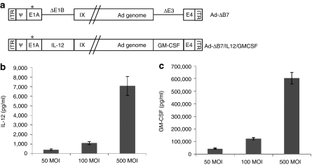
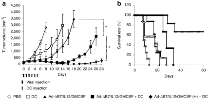
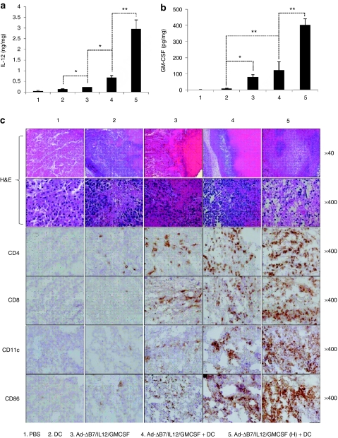
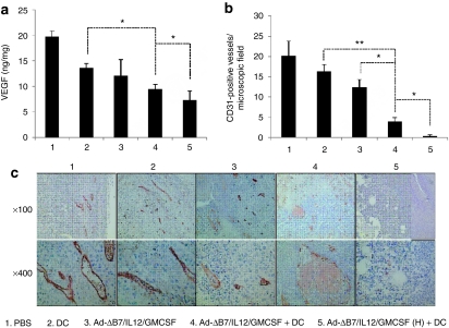
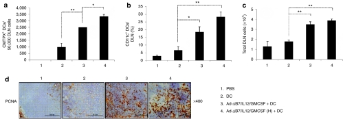
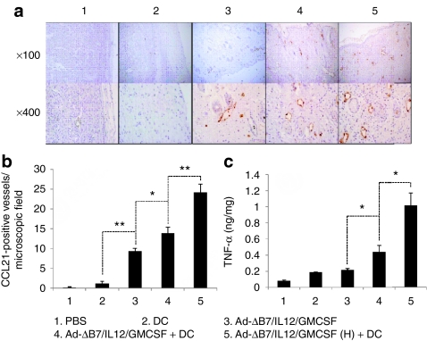
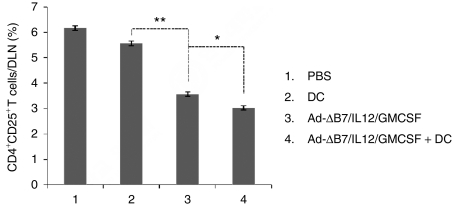
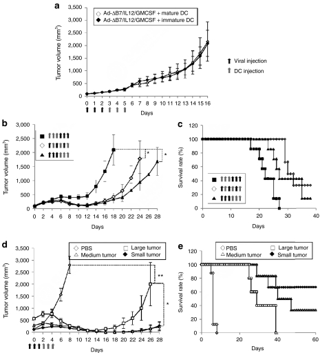
Dendritic cell (DC)-based vaccination is a promising strategy for cancer immunotherapy. However, clinical trials have indicated that immunosuppressive microenvironments induced by tumors profoundly suppress antitumor immunity and inhibit vaccine efficacy, resulting in insufficient reduction of tumor burdens. To overcome these obstacles and enhance the efficiency of DC vaccination, we generated interleukin (IL)-12- and granulocyte-macrophage colony-stimulating factor (GM-CSF)-coexpressing oncolytic adenovirus (Ad-ΔB7/IL12/GMCSF) as suitable therapeutic adjuvant to eliminate immune suppression and promote DC function. By treating tumors with Ad-ΔB7/IL12/GMCSF prior to DC vaccination, DCs elicited greater antitumor effects than in response to either treatment alone. DC migration to draining lymph nodes (DLNs) dramatically increased in mice treated with the combination therapy. This result was associated with upregulation of CC-chemokine ligand 21 (CCL21(+)) lymphatics in tumors treated with Ad-ΔB7/IL12/GMCSF. Moreover, the proportion of CD4(+)CD25(+) T-cells and vascular endothelial growth factor (VEGF) expression was decreased in mice treated with the combination therapy. Furthermore, combination therapy using immature DCs also showed effective antitumor effects when combined with Ad-ΔB7/IL12/GMCSF. The combination therapy had a remarkable therapeutic efficacy on large tumors. Taken together, oncolytic adenovirus coexpressing IL-12 and GM-CSF in combination with DC vaccination has synergistic antitumor effects and can act as a potent adjuvant for promoting and optimizing DC vaccination.


Figures

Figure 1
Figure 1
Characterization of oncolytic adenovirus coexpressing interleukin (IL)-12 and granulocyte-macrophage colony-stimulating factor (GM-CSF). (a) Schematic representation of the genomic structures of adenovirus (Ad) Ad-ΔB7 and Ad-ΔB7/IL12/GMCSF. Ad-ΔB7 contains mutated E1A (open star, mutation at Rb protein–binding site), but lacks E1B 19 and 55 kDa (ΔE1B), and E3 region (ΔE3); the murine IL-12 and murine GM-CSF were inserted into E1 and E3 region of Ad genome, respectively. The level of IL-12 (b) and GM-CSF (c) expression was confirmed in B16-F10 cells after infection with Ad-ΔB7/IL12/GMCSF at different MOIs. Cell culture supernatants were collected at 48 hour after infection, and the level of IL-12 and GM-CSF was quantified by conventional enzyme-linked immunosorbent assay kit. Data represent the mean ± SE of triplicate experiments, and similar results were obtained from at least three separate experiments. MOI, multiplicity of infection.
Figure 2
Figure 2
Antitumor effect of Ad-ΔB7/IL12/GMCSF in combination with dendritic cell (DCs). (a) Pre-established B16-F10 tumor was injected with phosphate-buffered saline (PBS) (open diamonds), DCs (open squares), Ad-ΔB7/IL12/GMCSF (filled triangles), Ad-ΔB7/IL12/GMCSF plus DCs (filled squares), or Ad-ΔB7/IL12/GMCSF (H; high-dose of Ad) plus DCs (filled diamonds). Tumor growth was monitored every day. (b) Survival percentage of tumor-bearing mice following treatment with Ad-ΔB7/IL12/GMCSF and/or DCs. Tumor size over 3,000 mm3 was regarded as death. Data points represent the mean ± SE. All results in this figure represent results of at least seven mice per group. *P < 0.05. GM-CSF, granulocyte-macrophage colony-stimulating factor; IL, interleukin.
Figure 3
Figure 3
Generation of antitumor immune response by Ad-ΔB7/IL12/GMCSF in combination with dendritic cell (DCs). The level of interleukin (IL)-12 (a) and granulocyte-macrophage colony-stimulating factor (GM-CSF) (b) expression in tumor tissues treated with phosphate-buffered saline (PBS), DCs, Ad-ΔB7/IL12/GMCSF, Ad-ΔB7/IL12/GMCSF plus DCs, or Ad-ΔB7/IL12/GMCSF (H; high-dose of Ad) plus DCs. (c) Histological and immunohistochemical analysis in tumor tissues treated with Ad-ΔB7/IL12/GMCSF and/or DCs. Tumor tissues were collected from mice at 3 days after final treatment, and paraffin section of tumor tissue was stained with hematoxylin and eosin (H&E) (top two rows, original magnification: ×40 and ×400). Immune cells infiltrated in tumor tissues were examined by anti-CD4 antibody (third row), anti-CD8 antibody (fourth row), anti-CD11c antibody (fifth row) and anti-CD86 antibody (bottom row). Original magnification: ×400. Data points represent the mean ± SE of at least three mice per group, and similar results were obtained from at least three separate experiments. *P < 0.05; **P < 0.01.
Figure 4
Figure 4
Decreased vascular endothelial growth factor (VEGF) expression and angiogenesis in tumors treated with combination therapy. Tumors were treated with phosphate-buffered saline (PBS), dendritic cells (DCs), Ad-ΔB7/IL12/GMCSF, Ad-ΔB7/IL12/GMCSF plus DCs, or Ad-ΔB7/IL12/GMCSF (H; high-dose of Ad) plus DCs, and harvested from mice at 3 days after final treatment. (a) The level of VEGF expression in tumor tissues was quantified by conventional enzyme-linked immunosorbent assay. *P < 0.05. (b) CD31+ microvessel immunohistochemistry in the tumor tissues treated with PBS, DC, Ad-ΔB7/IL12/GMCSF, Ad-ΔB7/IL12/GMCSF plus DCs, or Ad-ΔB7/IL12/GMCSF (H) plus DCs. Original magnification: ×100 and ×400. (c) The means of CD31+ vessel density in microscopic field for each treatment group (×200). Data points represent the mean ± SE of three mice per group, and similar results were obtained from at least two separate experiments. *P < 0.05; **P < 0.01. GM-CSF, granulocyte-macrophage colony-stimulating factor; IL, interleukin.
Figure 5
Figure 5
Dendritic cells (DC) migration to the draining lymph nodes (DLNs). Ex vivo generated DCs were labeled with CMTPX. Two days after final treatment, single cells were collected from DLNs and the migration was quantified by fluorescence-activated cell sorting analysis. (a) The number of CMTPX+ DCs in DLNs from mice treated with phosphate-buffered saline (PBS), DC, Ad-ΔB7/IL12/GMCSF plus DCs, or Ad-ΔB7/IL12/GMCSF (H; high-dose of Ad) plus DCs were quantified on a fluorescence-activated cell sorter, and data from 50,000 events were represented. (b) CD11c+ DCs in the DLNs from mice treated with Ad-ΔB7/IL12/GMCSF and/or DCs. (c) Total cell number of DLNs from different groups of mice. Data points represent the mean ± SE of triplicate experiments. All results in this figure represent results of at least three mice per group, and similar results were obtained from at least two separate experiments. *P < 0.05; **P < 0.01. (d) Immunohistochemical staining of proliferating cell nuclear antigen in draining lymph node tissues. Original magnification: ×400. GM-CSF, granulocyte-macrophage colony-stimulating factor; IL, interleukin.
Figure 6
Figure 6
Visualization and quantification of CCL21+ lymphatic vessels in tumors treated with Ad-ΔB7/IL12/GMCSF and/or dendritic cells (DCs). Tumors harvested from mice treated with phosphate-buffered saline (PBS), DC, Ad-ΔB7/IL12/GMCSF, Ad-ΔB7/IL12/GMCSF plus DCs, or Ad-ΔB7/IL12/GMCSF (H; high-dose of Ad) plus DCs were embedded in paraffin and sectioned. (a) Immunohistochemical staining of CCL21+ lymphatic vessels in tumor tissues. Original magnification: ×100 & ×400. (b) The means of CCL21+ lymphatic vessel in microscopic field (×200). (c) Tumor necrosis factor (TNF)-α expression in the tumor tissues was measured by conventional enzyme-linked immunosorbent assay. Data points represent the mean ± SE of triplicate experiments. All results in this figure represent results of at least three mice per group, and similar results were obtained from at least two separate experiments. *P < 0.05; **P < 0.01. GM-CSF, granulocyte-macrophage colony-stimulating factor; IL, interleukin.
Figure 7
Figure 7
CD4+CD25+ T-cells in draining lymph nodes (DLNs) from mice treated with Ad-ΔB7/IL12/GMCSF and/or dendritic cells (DCs). DLNs were collected from mice treated with phosphate-buffered saline (PBS), DC, Ad-ΔB7/IL12/GMCSF, Ad-ΔB7/IL12/GMCSF plus DCs, or Ad-ΔB7/IL12/GMCSF (H; high-dose of Ad) plus DCs. Proportion of CD4+CD25+ T-cells in the DLNs from different groups of mice were measured by fluorescence-activated cell sorting analysis. Data points represent the mean ± SE of three mice per group, and similar results were obtained from at least two separate experiments. *P < 0.05; **P < 0.01. GM-CSF, granulocyte-macrophage colony-stimulating factor; IL, interleukin.
Figure 8
Figure 8
Therapeutic efficacy of combination therapy based on the dendritic cells (DC) maturation, treatment schedule, and tumor size. (a) Tumor-bearing mice were treated with Ad-ΔB7/IL12/GMCSF in combination with mature or immature DCs. Similar antitumor effects were observed in combination therapies using mature or immature DCs. (b,c) Effect of the treatment schedule of oncolytic adenovirus and DCs on antitumor efficacy. Tumors were treated with Ad-ΔB7/IL12/GMCSF prior to DC vaccination (filled triangles), Ad-ΔB7/IL12/GMCSF and DCs on alternating days (open diamonds), or Ad-ΔB7/IL12/GMCSF after DC vaccination (filled squares). Tumor treatment with Ad-ΔB7/IL12/GMCSF prior to DC vaccination induced the most potent antitumor efficacy. (d,e) Antitumor effect of combination therapies on large tumors. High-dose combination therapy induced potent antitumor effect on large tumors. Data points represent the mean ± SE. All results in this figure represent results of at least six mice per group. *P < 0.05; **P < 0.01. GM-CSF, granulocyte-macrophage colony-stimulating factor; IL, interleukin.

Similar articles

Cited by

References

    1. Fong L., and, Engleman EG. Dendritic cells in cancer immunotherapy. Annu Rev Immunol. 2000;18:245–273. - PubMed
    1. Proudfoot O, Pouniotis D, Sheng KC, Loveland BE., and, Pietersz GA. Dendritic cell vaccination. Expert Rev Vaccines. 2007;6:617–633. - PubMed
    1. Asavaroengchai W, Kotera Y., and, Mulé JJ. Tumor lysate-pulsed dendritic cells can elicit an effective antitumor immune response during early lymphoid recovery. Proc Natl Acad Sci USA. 2002;99:931–936. - PMC - PubMed
    1. Nestle FO, Alijagic S, Gilliet M, Sun Y, Grabbe S, Dummer R.et al. (1998Vaccination of melanoma patients with peptide- or tumor lysate-pulsed dendritic cells Nat Med 4328–332. - PubMed
    1. Tjoa BA, Erickson SJ, Bowes VA, Ragde H, Kenny GM, Cobb OE.et al. (1997Follow-up evaluation of prostate cancer patients infused with autologous dendritic cells pulsed with PSMA peptides Prostate 32272–278. - PubMed

Publication types

MeSH terms