. 2011:2:270.
doi: 10.1038/ncomms1270.

Annexin-A5 assembled into two-dimensional arrays promotes cell membrane repair

Affiliations
Free PMC article

Annexin-A5 assembled into two-dimensional arrays promotes cell membrane repair

Anthony Bouter et al. Nat Commun. 2011.
Free PMC article

Abstract

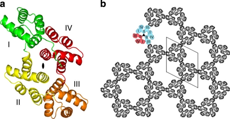
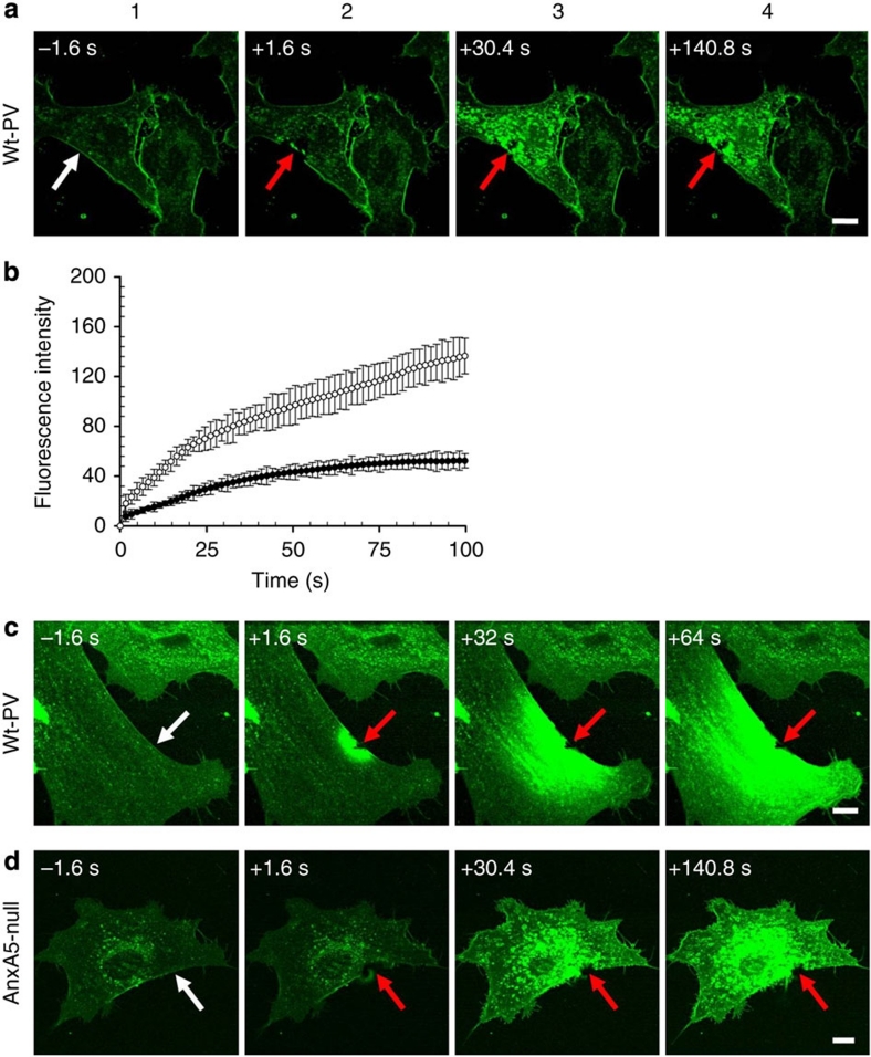
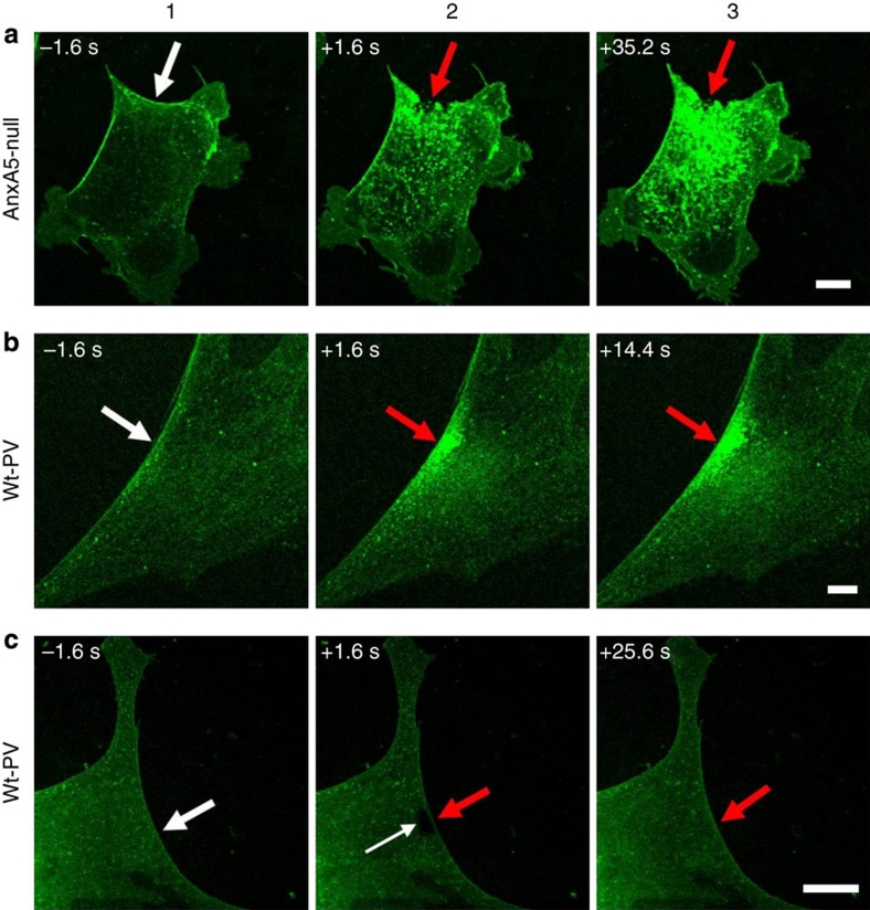
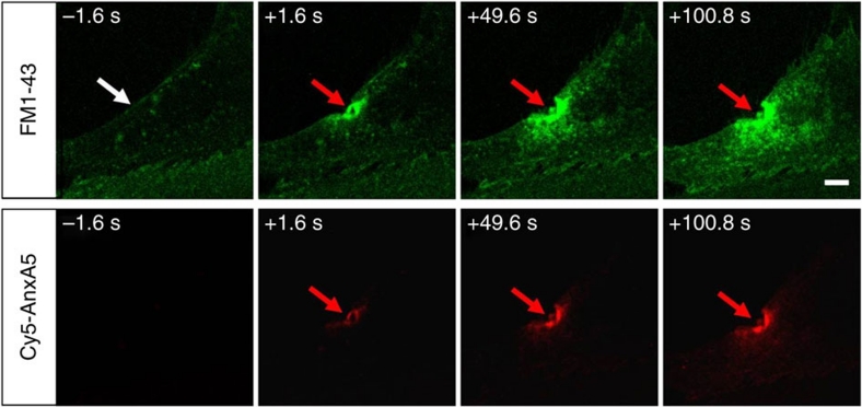
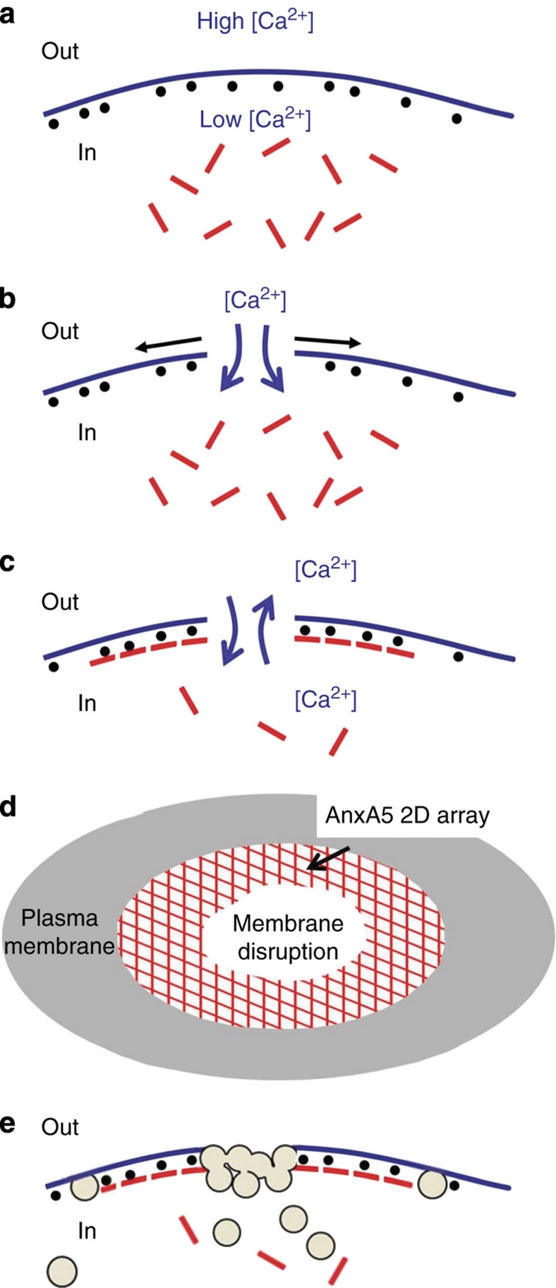
Eukaryotic cells possess a universal repair machinery that ensures rapid resealing of plasma membrane disruptions. Before resealing, the torn membrane is submitted to considerable tension, which functions to expand the disruption. Here we show that annexin-A5 (AnxA5), a protein that self-assembles into two-dimensional (2D) arrays on membranes upon Ca(2+) activation, promotes membrane repair. Compared with wild-type mouse perivascular cells, AnxA5-null cells exhibit a severe membrane repair defect. Membrane repair in AnxA5-null cells is rescued by addition of AnxA5, which binds exclusively to disrupted membrane areas. In contrast, an AnxA5 mutant that lacks the ability of forming 2D arrays is unable to promote membrane repair. We propose that AnxA5 participates in a previously unrecognized step of the membrane repair process: triggered by the local influx of Ca(2+), AnxA5 proteins bind to torn membrane edges and form a 2D array, which prevents wound expansion and promotes membrane resealing.


Figures

Figure 1
Figure 1. Structure of soluble and membrane-bound AnxA5.
(a) View of an AnxA5 molecule from the concave side of the molecule. AnxA5, the smallest annexin, is made up of four α-helical domains, numbered I–IV, which form the conserved membrane-binding core of annexins. The molecule is observed along the pseudo twofold symmetry axis (dark ellipse), which relates module (I+IV) to module (II+III). This orientation corresponds almost to that of membrane-bound AnxA5 (refs 2758). This figure was created with PyMOL (http://pymol.org/) from PDB code 1AVR. (b) Projection view of a 2D crystal of membrane-bound AnxA5 (adapted from ref. 35). On membrane binding, AnxA5 molecules self-assemble into trimers and 2D crystals of trimers with various packing arrangements. Panel b represents the main 2D crystalline arrangement formed by AnxA5 trimers, which has the symmetry of the two-sided plane group p6 (ref. 35). A hexagonal unit cell is represented, with 17.7-nm unit cell side. An AnxA5 trimer is coloured, with one red and two blue monomers.
Figure 2
Figure 2. Responses of wt-PV and AnxA5-null PV cells to 160-mW infrared irradiation.
(a) Sequences of images showing the response of a wt-PV cell to 160-mW infrared irradiation in the presence of 2-mM Ca2+. In this panel, as well as in panels c and d, the area of membrane irradiation is marked with a white arrow before irradiation and a red arrow after irradiation. Image frames 1 and 2 were recorded 1.6 s before and 1.6 s after irradiation, respectively; frames 3–4 were recorded 30.4 and 140.8 s after irradiation, respectively, as indicated. (b) Kinetics of FM1-43 fluorescence intensity increase for wt-PV cells (filled circles) and AnxA5-null PV cells (empty circles) after membrane damage at 160-mW infrared irradiation in the presence of 2-mM Ca2+. Data represent the fluorescence intensity integrated over whole cell sections, averaged for 10 cells (±s.d.). For wt-PV cells, the fluorescence intensity reaches a plateau after ∼90 s. For AnxA5-null PV cells, the fluorescence intensity increases continuously and is significantly larger than that for wt-PV cells, about ×3 larger at 100 s. (c) Sequence of images showing the response of a wt-PV cell to 160-mW infrared irradiation in the presence of 1-mM EGTA. Before irradiation, cells were washed three times in DPBS containing no CaCl2, then incubated in DPBS supplemented with 1-mM EGTA for 5 min. (d) Sequence of images showing the response of an AnxA5-null PV cell to 160-mW infrared irradiation in the presence of 2-mM Ca2+. Scale bars, 10 μm.
Figure 3
Figure 3. Responses of wt-PV and AnxA5-null PV cells to 80-mW infrared irradiation.
(a) Representative image series showing the response of an AnxA5-null PV cell to 80-mW infrared irradiation. The cell presents a macroscopic membrane rupture at the irradiated area and a large increase of cytoplasmic fluorescence intensity, as observed at 160 mW. (b,c) Image series showing two types of response for wt-PV cells irradiated at 80 mW. (b) A minor fraction (about 30%) of wt-PV cells presents an increase in cytoplasmic fluorescence intensity, yet no macroscopic membrane rupture. (c) The major fraction (about 70%) of wt-PV cells shows no increase in cytoplasmic fluorescence intensity. The areas of membrane irradiation are marked with a white arrow before irradiation and a red arrow after irradiation. Scale bars, 10 μm.
Figure 4
Figure 4. Binding of Cy5-AnxA5 to injured membranes.
Simultaneous recording of FM1-43 (top) and Cy5-AnxA5 (bottom) fluorescence of a wt-PV cell submitted to 160-mW infrared-induced membrane rupture. Cy5-AnxA5 binds exclusively at the level of the injured membrane, colocalizing initially with FM1-43 dyes. The area of membrane irradiation is marked with a white arrow before irradiation and a red arrow after irradiation. Scale bar, 10 μm.
Figure 5
Figure 5. Structure and packing of AnxA5 and mtT-AnxA5 in three-dimensional crystals.
(a, b) Structure of trimers of recombinant rat AnxA5 (a) and monomer–monomer interface of a trimer (b) in R3 crystals (created from PDB code 1a8a). (a) The intramolecular pseudo twofold axes, which relate (I+IV) and (III+II) modules (indicated by crosses in each monomer of the bottom trimer) lie almost parallel (12°) to the threefold symmetry axes (black triangles) normal to the (a, b) plane. (b) Detailed structure of the monomer–monomer interface of an AnxA5 trimer, viewed along the arrow orientation in (a). A–E refer to AnxA5 α-helices, according to annexin nomenclature. Several pairs of charged amino acids located at distances close enough to form salt bridges are indicated in ball-and-stick representation, with positively and negatively charged residues in blue and red, respectively. (c) Packing of mtT-AnxA5 molecules in high Ca2+ content 3D crystals, along the b monoclinic axis. MtT-AnxA5 molecules crystallize in the P21 space group, with lattice parameters a=51.108 Å, b=67.184 Å, c=112.313 Å, β=94.79°. The asymmetric unit contains two protein molecules related by a non-crystallographic 2ɛ screw axis parallel to the a axis, with a non-fractional pitch of ɛ=4.7 Å. The positions of two 2ɛ screw axes are indicated. MtT-AnxA5 molecules are oriented in the a, c plane with the intramolecular pseudo twofold axis (indicated by a cross in the AnxA5 monomer with I–IV numbering) lying almost normal (12°) to the a, c plane. This orientation is thus similar to that of native AnxA5 molecules shown in Figure 5a. Side chains of residues involved in salt-bridge interactions in AnxA5 trimers are shown as blue sticks. None of the residues participates in intermolecular contacts, with the exception of residue E16, which interacts through a Ca2+-mediated salt bridge with a neighbouring molecule along the b axis.
Figure 6
Figure 6. Binding of mtT-AnxA5 to model membranes and infrared-injured cells.
(a) Comparison of the binding of native AnxA5 (blue curve) and mtT-AnxA5 (red curve) to a supported lipid bilayer made of DOPC/DOPS (7:3, w/w), by QCM-D. The formation of a supported lipid bilayer by deposition of small unilamellar vesicles on silica-coated quartz crystals takes place before t=0 mn and is not shown here. At t=0 min, a solution of 5-μg ml−1 AnxA5 or mtT-AnxA5 in HBS-Ca, pH 7.4, was injected at 150 μl min−1. The kinetics of adsorption and the adsorbed masses at saturation are almost identical for both proteins. The small difference observed between the kinetics of binding of native AnxA5 and mT-AnxA5 is equivalent to a difference in protein concentration of <1 μg ml−1, that is, 20% of nominal protein concentration, which is the level of accuracy of standard protein concentration assays. (b, c) Simultaneous recording of Cy5-mtT-AnxA5 (b) and FM1-43 (c) fluorescence of a wt-PV cell 1.6 s after 160-mW infrared-induced membrane rupture. The red arrows indicate the area of membrane irradiation. Scale bar, 10 μm.
Figure 7
Figure 7. Model of cell membrane repair.
(a) Intact cell membrane. The extracellular (Out) and intracellular (In) milieus differ by their high versus low Ca2+ concentrations, and the absence versus presence of AnxA5 (red rods) and PS (black spheres), respectively. (b) Local rupture of a cell membrane. Forces resulting from membrane tension tend to expand the tear laterally (black arrows). (c) The formation of an AnxA5 2D array at the torn membrane stops wound expansion. On membrane rupture, a microenvironment is formed in which the intracellular and extracellular milieus mix, providing optimal conditions for the formation of tight complexes between Ca2+, PS and AnxA5 (ref. 44). AnxA5 molecules bind to PS molecules exposed at ruptured membrane edges, form trimers and 2D arrays. These AnxA5 2D self-assemblies stabilize the membrane and stop expansion of the tear. (d) Top view of a membrane disruption with an AnxA5 2D array surrounding the tear. (e) Membrane resealing. Cytoplasmic vesicles are recruited at the ruptured membrane area and fuse by exocytosis with the plasma membrane, either directly as single vesicles or as a patch formed by homotypic fusion of intracellular vesicles. The resulting increase in membrane surface and decrease in membrane tension lead to membrane resealing.

References

    1. McNeil P. L. & Steinhardt R. A. Plasma membrane disruption: repair, prevention, adaptation. Annu. Rev. Cell Dev. Biol. 19, 697–731 (2003). - PubMed
    1. Glover L. & Brown R. H. Dysferlin in membrane trafficking and patch repair. Traffic 8, 785–794 (2007). - PubMed
    1. McNeil P. L. & Kirchhausen T. An emergency response team for membrane repair. Nat. Rev. Mol. Cell Biol. 6, 499–505 (2005). - PubMed
    1. Steinhardt R. A., Bi G. & Alderton J. M. Cell membrane resealing by a vesicular mechanism similar to neurotransmitter release. Science 263, 390–393 (1994). - PubMed
    1. Sheetz M. P. & Dai J. Modulation of membrane dynamics and cell motility by membrane tension. Trends Cell Biol. 6, 85–89 (1996). - PubMed

Publication types

Associated data