. 2011 Apr;32(4):456-64.
doi: 10.1038/aps.2011.9.

The vasorelaxing effect of hydrogen sulfide on isolated rat aortic rings versus pulmonary artery rings

Affiliations

The vasorelaxing effect of hydrogen sulfide on isolated rat aortic rings versus pulmonary artery rings

Yan Sun et al. Acta Pharmacol Sin. 2011 Apr.

Abstract

Aim: To compare the vasorelaxing effects of hydrogen sulfide (H(2)S) on isolated aortic and pulmonary artery rings and to determine their action mechanisms.

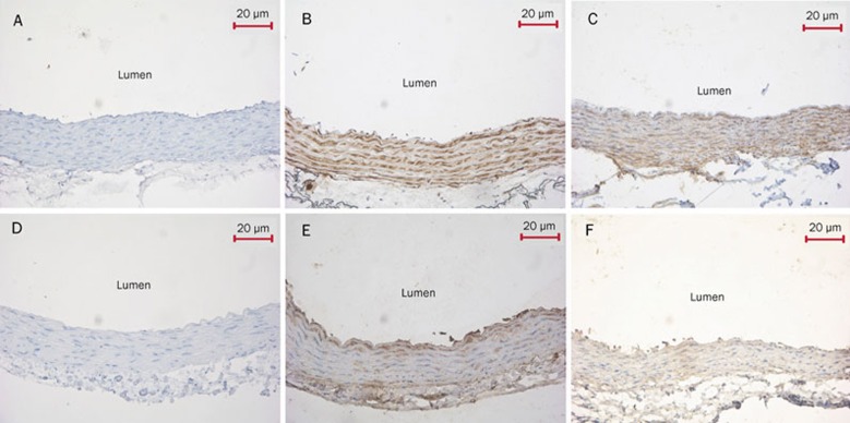
Methods: H(2)S-induced vasorelaxation of isolated rat aortic versus pulmonary artery rings under 95% O(2) and 5% CO(2) was analyzed. The expression of cystathinonine gamma-lyase (CSE), cystathionine beta synthase (CBS), 3-mercaptopyruvate sulfurtransferase (3MST), SUR2B and Kir6.1 was examined.

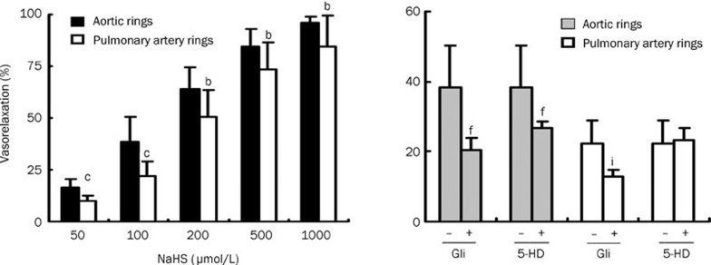
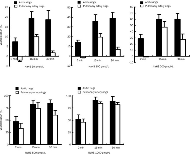
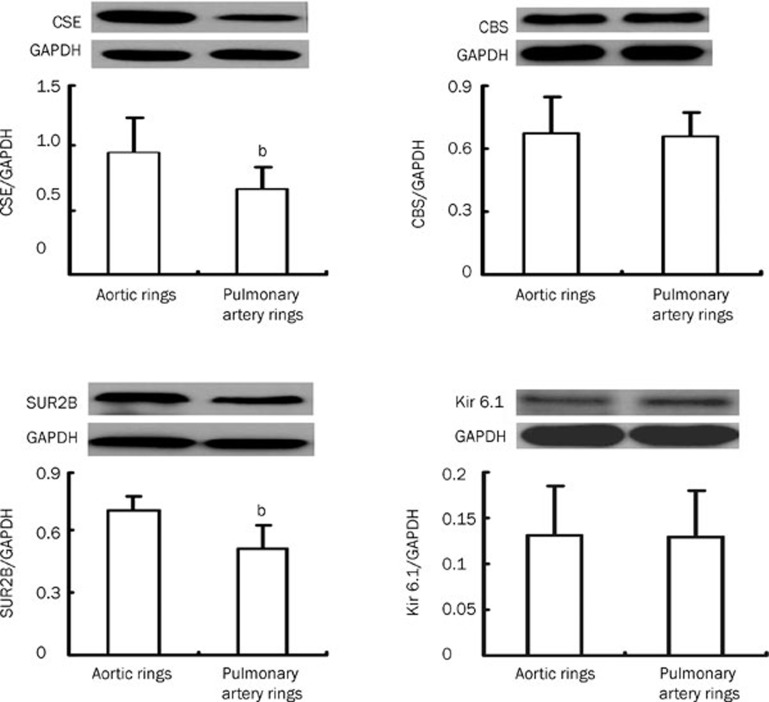
Results: NaHS caused vasorelaxation of rat aortic and pulmonary artery rings in a dose-dependent manner. NaHS dilated aortic rings to a greater extent (16.4%, 38.4%, 64.1%, 84.3%, and 95.9% at concentrations of 50, 100, 200, 500, and 1000 μmol/L, respectively) than pulmonary artery rings (10.1%, 22.2%, 50.6%, 73.6%, and 84.6% at concentrations of 50, 100, 200, 500 and 1000 μmol/L, respectively). The EC(50) of the vasorelaxant effect for aortic rings was 152.17 μmol/L, whereas the EC(50) for pulmonary artery rings was 233.65 μmol/L. The vasorelaxing effect of H(2)S was markedly blocked b y cellular and mitochondrial membrane K(ATP) channel blockers in aortic rings (P<0.01). In contrast, only the cellular membrane K(ATP) channel blocker inhibited H(2)S-induced vasorelaxation in pulmonary artery rings. SUR2B mRNA and protein expression was higher in aortic rings than in pulmonary artery rings. Cystathinonine gamma-lyase (CSE) but not cystathionine beta synthase (CBS) expression in aortic rings was higher than in pulmonary artery rings. 3-Mercapto pyruvate sulfurtransferase (3MST) mRNA was lower in aortic rings than in pulmonary artery rings.

Conclusion: The vasorelaxing effect of H(2)S on isolated aortic rings was more pronounced than the effect on pulmonary artery rings at specific concentrations, which might be associated with increased expression of the K(ATP) channel subunit SUR2B.


Figures

Figure 1
Figure 1
The maximum relaxation response of aortic and pulmonary artery rings to different concentrations of NaHS in rats, and the effect of a KATP channel blocker on the vasorelaxing effect of hydrogen sulfide on aortic and pulmonary artery rings (n=10). bP<0.05, cP<0.01 compared to aortic rings. fP<0.01 compared to aortic rings without giving KATP channel blocker. iP<0.01 compared to pulmonary artery rings without giving KATP channel blocker. Gli: glibenclamide, 5-HD: 5-hydroxydecanoate.
Figure 2
Figure 2
The different vasoactive response of aortic and pulmonary artery rings to different concentrations of NaHS in rats at the different time points.
Figure 3
Figure 3
Immunohistochemical analysis of CSE expression in aortic and pulmonary artery rings (DAB×200). (A) The aortic ring negative control was processed without CSE primary antibody. The thickness of the inner elastic layer was uniform and the structure of smooth muscle cells was normal. (B) The aortic ring was processed with CSE antibody. CSE protein was strongly expressed in the inner membrane and tunica media vasorum. The brown granules in aortic smooth muscle cells and endothelial cells were defined as positive signals. (C) The pulmonary artery ring was processed with CSE antibody. The brown granules were observed in pulmonary artery smooth muscle cells and endothelial cells. CSE, cystathinonine gamma-lyase.
Figure 4
Figure 4
CSE, CBS, SUR2B and Kir6.1 expression in aortic and pulmonary artery rings as detected by Western blotting (n=10, mean±SD). bP<0.05 compared to aortic rings. CSE, cystathinonine gamma-lyase; CBS, cystathionine beta synthase; SUR2B, a KATP channel subunit; Kir6.1, a KATP channel subunit.
Figure 5
Figure 5
Expression of SUR2B, Kir6.1 and 3MST by real-time PCR (n=7, mean±SD). bP<0.05 compared to aortic rings. SUR2B, a KATP channel subunit. Kir6.1, a KATP channel subunit. 3MPST, 3-mercaptopyruvate sulfurtransferase.
Figure 6
Figure 6
Immunohistochemical analysis of SUR2B and Kir6.1 expression in aortic and pulmonary artery rings (DAB×200). (A) The aortic ring negative control was processed without SUR2B primary antibody. This control had normal smooth muscle cell and endothelial cell structure without brown granules. (B) The aortic ring was treated with SUR2B antibody. SUR2B protein was strongly expressed in the inner membrane and tunica media vasorum. The presence of the brown granules in aortic smooth muscle cells and endothelial cells was defined as positive signals. (C) The pulmonary artery ring was processed with SUR2B antibody. Brown granules were observed in pulmonary artery smooth muscle cells and endothelial cells. (D) The aortic ring negative control was treated without Kir6.1 primary antibody. The structure of smooth muscle cells and endothelial cells was normal and had no brown granules. (E) The aortic ring was treated with Kir6.1 antibody. Brown granules were strongly expressed in the medial layer of aortic ring. (F) The pulmonary artery ring was processed with Kir6.1 antibody. The brown granules were observed in pulmonary artery smooth muscle cells and endothelial cells. SUR2B, a KATP channel subunit. Kir6.1, a KATP channel subunit.

Similar articles

Cited by

References

    1. Aaronson PI, Robertson TP, Knock GA, Becker S, Lewis TH, Snetkov V, et al. Hypoxic pulmonary vasoconstriction: mechanisms and controversies. J Physiol. 2006;570:53–8. - PMC - PubMed
    1. Gray GA, Webb DJ. The endothelin system and its potential as a therapeutic target in cardiovascular disease. Pharmacol Ther. 1996;72:109–48. - PubMed
    1. Sata M, Fukuda D. Crucial role of renin-angiotensin system in the pathogenesis of atherosclerosis. J Med Invest. 2010;57:12–25. - PubMed
    1. Lipworth BJ, Dagg KD. Vasoconstrictor effects of angiotensin II on the pulmonary vascular bed. Chest. 1994;105:1360–4. - PubMed
    1. Olson KR, Dombkowski RA, Russell MJ, Doellman MM, Head SK, Whitfield NL, et al. Hydrogen sulfide as an oxygen sensor/transducer in vertebrate hypoxic vasoconstriction and hypoxic vasodilation. J Exp Biol. 2006;209:4011–23. - PubMed

Publication types