. 2011 Sep;31(9):1919-29.
doi: 10.1038/jcbfm.2011.42. Epub 2011 Apr 6.

Long-lasting protection in brain trauma by endotoxin preconditioning

Affiliations

Long-lasting protection in brain trauma by endotoxin preconditioning

Luca Longhi et al. J Cereb Blood Flow Metab. 2011 Sep.

Abstract

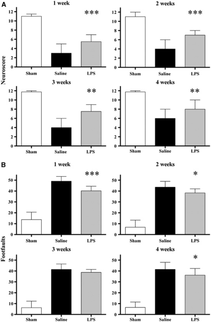
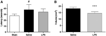
We investigated the occurrence of endotoxin (lipopolysaccharide, LPS) preconditioning in traumatic brain injury (TBI), evaluating the time window of LPS-induced protection, its persistence, and the associated molecular mechanisms. Mice received 0.1 mg/kg LPS or saline intraperitoneally and subsequently TBI (by controlled cortical impact brain injury) at various time intervals. Mice receiving LPS 3, 5, or 7 days before TBI showed attenuated motor deficits at 1 week after injury compared with mice receiving saline. Those receiving LPS 5 days before injury had also a reduced contusion volume (7.9±1.3 versus 12±2.3 mm(3)) and decreased cell death. One month after injury, the protective effect of LPS on contusion volume (14.5±1.2 versus 18.2±1.2 mm(3)) and neurologic function was still present. Traumatic brain injury increased glial fibrillary acidic protein, CD11b, CD68, tumor necrosis factor-α, interleukin (IL)-10, and IL-6 mRNA expression 24 hours after injury. Lipopolysaccharide administered 5 (but not 9) days before injury increased the expression of CD11b (233%) and of interferon β (500%) in uninjured mice, while it reduced the expression of CD68 (by 46%) and increased that of IL-6 (by 52%) in injured mice. Lipopolysaccharide preconditioning conferred a long-lasting neuroprotection after TBI, which was associated with a modulation of microglia/macrophages activity and cytokine production.


Figures

Figure 1
Figure 1
Time window of lipopolysaccharide (LPS) preconditioning. (A) Neuroscore at 1 week after injury after either saline or LPS administration at various time points before traumatic brain injury (TBI). Data are reported as median+range (n=12). Kruskall–Wallis analysis of variance (ANOVA) (P<0.01) followed by Mann–Whitney U-test for individual comparisons. ##P<0.01 compared with saline and LPS at 1 and 9 days interval. (B) Representative coronal sections taken at approximately −1.8 mm from bregma, at 1 week after injury after either saline or LPS administration at various time points before TBI, showing the extent of cortical tissue loss and the atrophy of the ipsilateral hippocampus. (C) Quantification of contusion volume in experimental groups as in (B). Data are reported as mean+s.d. (n=6). ANOVA: P<0.05 followed by Newman–Keuls multiple comparisons. #P<0.05 compared with saline. (D) Quantification of transferase-mediated dUTP nick end labeling (TUNEL)-positive cells in the injured cortex of mice receiving saline or LPS 5 days before TBI, at 1 week after injury. Data are reported as mean+s.d. (n=6). **P<0.01 compared with saline.
Figure 2
Figure 2
Long-term effects of lipopolysaccharide (LPS) preconditioning. Neuromotor function over 4 weeks after injury after either saline or LPS administration 5 days before traumatic brain injury (TBI). (A) Neuroscore: data are reported as median+range (sham: n=4; brain injured: n=12). Kruskall–Wallis analysis of variance (ANOVA) followed by Mann–Whitney U-test for individual comparison. (B) Beam walk: data are reported as mean+s.d. ANOVA followed by Newman–Keuls post hoc test. ***P<0.001,**P<0.01, and *P<0.05 compared with saline.
Figure 3
Figure 3
Long-term effects of lipopolysaccharide (LPS) preconditioning. (A) Cognitive function: latencies at 4 weeks after injury (average of 24 trials over 3 days) in mice receiving either saline or LPS 5 days before traumatic brain injury (TBI). Data are reported as mean+s.d. (sham: n=4; brain injured: n=12). Analysis of variance (ANOVA): P<0.05 followed by Newman–Keuls post hoc test. #P<0.05 compared with sham. (B) Histopathology: quantification of contusion volume at 5 weeks after injury in mice receiving either saline or LPS 5 days before TBI. Data are reported as mean+s.d. (n=6). ***P<0.001 compared with saline.
Figure 4
Figure 4
Gene expression. (A) mRNA expression in the injured cortex at 24 hours in sham or injured mice receiving either saline or lipopolysaccharide (LPS) 5 days before traumatic brain injury (TBI). Data are expressed as fold of induction compared with the sham-saline group. Data are reported as mean+s.d. (n=8). Two-way analysis of variance (ANOVA) followed by Bonferroni post hoc test: overall interaction P<0.01 for CD11b, CD68, and interleukin (IL)-6. **P<0.01, *P<0.05 LPS, compared with saline. (B) mRNA expression in the injured cortex at 24 hours in sham or injured mice receiving either saline or LPS 9 days before TBI. Data are reported as mean+s.d. (n=8). IFN, interferon.
Figure 5
Figure 5
Immunohistochemistry. Representative micrographs of CD11b (A) and CD68 (C) staining in the injured cortex of mice receiving either saline or lipopolysaccharide (LPS) 5 days before traumatic brain injury (TBI), at 1 week after injury. Bar: 10 μm. Quantification of microglia/macrophages activation by anti-CD11b immunostaining and of phagocytic activity by anti-CD68 immunostaining is shown in graphs (B, D), respectively. Data are expressed as percentage of stained area over the total sampled area and reported as mean+s.d. of 33 frames/mouse (n=6). **P<0.01 compared with saline.

References

    1. Ahmed SH, He YY, Nassief A, Xu J, Xu XM, Hsu CY, Faraci FM. Effects of lipopolysaccharide priming on acute ischemic brain injury. Stroke. 2000;31:193–199. - PubMed
    1. Assaf Y, Holokovsky A, Berman E, Shapira Y, Shohami E, Cohen Y. Diffusion and perfusion magnetic resonance imaging following closed head injury in rats. J Neurotrauma. 1999;16:1165–1176. - PubMed
    1. Bastide M, Gele P, Petrault O, Pu Q, Caliez A, Robin E, Deplanque D, Duriez P, Bordet R. Delayed cerebrovascular protective effect of lipopolysaccharide in parallel to brain ischemic tolerance. J Cereb Blood Flow Metab. 2003;23:399–405. - PubMed
    1. Botker HE, Kharbanda R, Schmidt MR, Bottcher M, Kaltoft AK, Terkelsen CJ, Munk K, Andersen NH, Hansen TM, Trautner S, Lassen JF, Christiansen EH, Krusell LR, Kristensen SD, Thuesen L, Nielsen SS, Rehling M, Sorensen HT, Redington AN, Nielsen TT. Remote ischaemic conditioning before hospital admission, as a complement to angioplasty, and effect on myocardial salvage in patients with acute myocardial infarction: a randomised trial. Lancet. 2010;375:727–734. - PubMed
    1. Capone C, Frigerio S, Fumagalli S, Gelati M, Principato MC, Storini C, Montinaro M, Kraftsik R, De Curtis M, Parati E, De Simoni MG. Neurosphere-derived cells exert a neuroprotective action by changing the ischemic microenvironment. PLoS One. 2007;2:e373. - PMC - PubMed

Publication types