

Glycoprotein 2 (GP2): grabbing the FimH bacteria into M cells for mucosal immunity
- PMID: 21468225
- PMCID: PMC3056108
- DOI: 10.4161/gmic.1.6.14078
Glycoprotein 2 (GP2): grabbing the FimH bacteria into M cells for mucosal immunity
Abstract
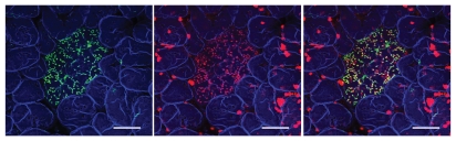
Membranous (M) cells are specialized epithelial antigen-transporting cells scattered in the follicle-associated epithelium covering the gut lymphoid follicles such as Peyer's patches. Although the importance of M cells as a main portal for luminal antigens has long been recognized, molecular mechanisms for M-cell antigen uptake has remained largely elusive. We have recently found that glycoprotein 2 (GP2) is exclusively expressed on M cells among intestinal epithelial cells and serves as an uptake receptor for a subset of commensal and pathogenic bacteria. GP2 interacts with FimH, a major component of the type 1 pilus on the outer membrane of a subset of gram-negative enterobacilli such as E. coli and Salmonella enterica. Furthermore, GP2-FimH interaction is necessary for efficient uptake of FimH(+) bacteria by M cells and subsequent bacteria-specific mucosal immune responses. Pancreatic GP2 may also be involved in innate immunity by 'opsonization' of FimH(+) bacteria to facilitate their egestion in feces as well as translocation across the intestinal epithelium.
Keywords: Escherichia coli; FimH; IgA; M cells; Salmonella typhimurium; antigen uptake; bacteria; mucosal immunity; type I pili.
© 2010 Landes Bioscience
Figures
Comment on
-
Uptake through glycoprotein 2 of FimH(+) bacteria by M cells initiates mucosal immune response.Nature. 2009 Nov 12;462(7270):226-30. doi: 10.1038/nature08529. Nature. 2009. PMID: 19907495
Similar articles
-
Uptake through glycoprotein 2 of FimH(+) bacteria by M cells initiates mucosal immune response.Nature. 2009 Nov 12;462(7270):226-30. doi: 10.1038/nature08529. Nature. 2009. PMID: 19907495
-
Application of a mouse ligated Peyer’s patch intestinal loop assay to evaluate bacterial uptake by M cells.J Vis Exp. 2011 Dec 17;(58):3225. doi: 10.3791/3225. J Vis Exp. 2011. PMID: 22215009 Free PMC article.
-
A novel mucosal vaccine targeting Peyer's patch M cells induces protective antigen-specific IgA responses.Int Immunol. 2014 Nov;26(11):619-25. doi: 10.1093/intimm/dxu061. Epub 2014 Jun 7. Int Immunol. 2014. PMID: 24908678
-
Molecular insights into the mechanisms of M-cell differentiation and transcytosis in the mucosa-associated lymphoid tissues.Anat Sci Int. 2018 Jan;93(1):23-34. doi: 10.1007/s12565-017-0418-6. Epub 2017 Nov 2. Anat Sci Int. 2018. PMID: 29098649 Review.
-
Roles of M cells in infection and mucosal vaccines.Hum Vaccin Immunother. 2014;10(12):3544-51. doi: 10.4161/hv.36174. Hum Vaccin Immunother. 2014. PMID: 25483705 Free PMC article. Review.
Cited by
-
Genome-wide association meta-analysis identifies GP2 gene risk variants for pancreatic cancer.Nat Commun. 2020 Jun 24;11(1):3175. doi: 10.1038/s41467-020-16711-w. Nat Commun. 2020. PMID: 32581250 Free PMC article.
-
Recent Trends in Non-Invasive Methods of Diagnosis and Evaluation of Inflammatory Bowel Disease: A Short Review.Int J Mol Sci. 2024 Feb 8;25(4):2077. doi: 10.3390/ijms25042077. Int J Mol Sci. 2024. PMID: 38396754 Free PMC article. Review.
-
Oral Administration of Cancer Vaccines: Challenges and Future Perspectives.Vaccines (Basel). 2023 Dec 26;12(1):26. doi: 10.3390/vaccines12010026. Vaccines (Basel). 2023. PMID: 38250839 Free PMC article. Review.
-
Serological markers of inflammatory bowel disease.Biochem Med (Zagreb). 2013;23(1):28-42. doi: 10.11613/bm.2013.006. Biochem Med (Zagreb). 2013. PMID: 23457764 Free PMC article. Review.
-
Comparative Study of Aryl O-, C-, and S-Mannopyranosides as Potential Adhesion Inhibitors toward Uropathogenic E. coli FimH.Molecules. 2019 Oct 2;24(19):3566. doi: 10.3390/molecules24193566. Molecules. 2019. PMID: 31581627 Free PMC article.
References
-
- Owen RL. Uptake and transport of intestinal macromolecules and microorganisms by M cells in Peyer's patches—a personal and historical perspective. Semin Immunol. 1999;11:157–163. - PubMed
-
- Kraehenbuhl JP, Neutra MR. Epithelial M cells: differentiation and function. Annu Rev Cell Dev Biol. 2000;16:301–332. - PubMed
-
- Kato T, Owen RL. Structure and function of intestinal mucosal epithelium. In: Mestecky J, Lamm ME, McGhee JR, Bienenstock J, Mayer L, Strober W, editors. Mucosal Immunology. Elsevier Academic Press; 2005. pp. 131–151.
-
- Kumagai K. Über den Resorptionsvergang der corpusculären Bestandteile im Darm. Kekkaku-Zassi. 1922;4:429–431. (Ger).
-
- Bockman DE, Cooper MD. Pinocytosis by epithelium associated with lymphoid follicles in the bursa of Fabricius, appendix and Peyer's patches. An electron microscopic study. Am J Anat. 1973;136:455–477. - PubMed
Publication types
MeSH terms
Substances
LinkOut - more resources
Full Text Sources
Other Literature Sources
Miscellaneous