

Successful ablation of a left-sided accessory pathway in a patient with coronary sinus atresia and arteriovenous fistula: clinical and developmental insights
- PMID: 21468248
- PMCID: PMC3063614
Successful ablation of a left-sided accessory pathway in a patient with coronary sinus atresia and arteriovenous fistula: clinical and developmental insights
Abstract
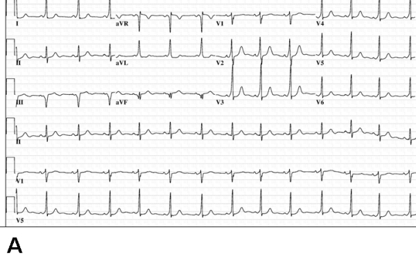
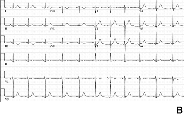
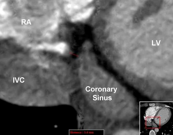
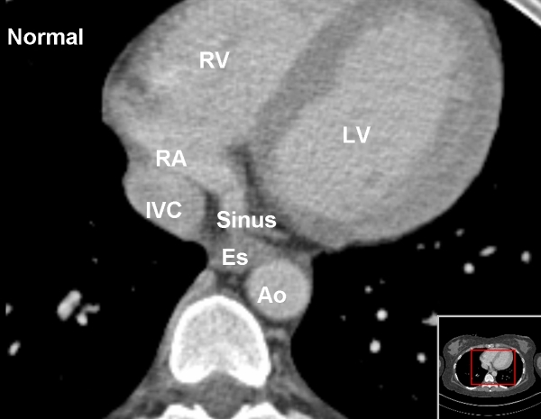
Background and objectives: While radiofrequency ablation catheter ablation of accessory pathways is generally safe and effective, anatomic variants can cause considerable challenges in effecting cure. Our objective was to use an unusual case where coronary sinus was absent and arterial venous fistula was present and a left-sided pathway required mapping and ablation to develop a framework to approach difficult cases.
Method: A detailed literature search and review of contemporary cardiac embryology was undertaken to attempt and to explain a common developmental anomaly. Adjunctive approaches during the ablation procedure, including intracardiac ultrasound, were used to guide mapping and ablation despite the lack of coronary sinus access.
Results: The accessory pathway was successfully ablated using a transseptal approach and intracardiac ultrasound guided mapping of the mitral annulus. A potential common mechanism to explain the apparently disparate anatomic variants in this patient was formulated.
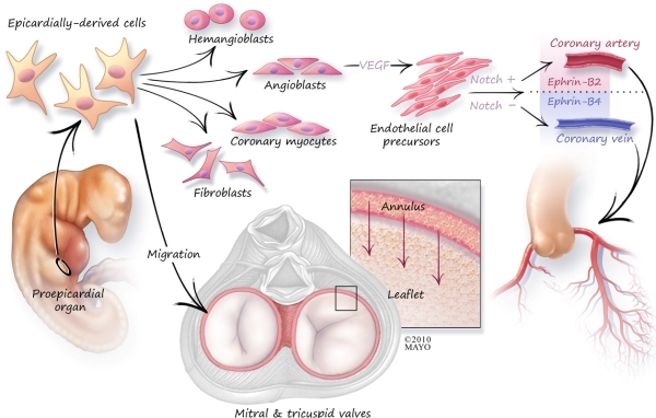
Conclusions: Cardiac conduction development is complex and accessory pathway conduction may occur in the setting of arteriovenous anomalies thus providing insights as to the cause of WPW syndrome. Successful mapping and targeted ablation of left-sided pathways may be accomplished even when coronary sinus access is not possible.
Keywords: Coronary sinus; accessory pathway; coronary AV fistula; coronary atresia; epicardially-derived cells.
Figures
References
-
- Kim J. Left-sided accessory pathway with ostial atresia of the coronary sinus: a case report. Pacing Clin Electrophysiol. 2008;31:129. - PubMed
-
- Kirby M. Cardiac Development. New York, NY: Oxford University Press; 2007.
-
- Arad M, et al. Transgenic mice overexpressing mutant PRKAG2 define the cause of Wolff-Parkinson-White syndrome in glycogen storage cardiomyopathy. Circulation. 2003;107:2850. - PubMed
-
- Hahurij ND, et al. Accessory atrioventricular myocardial connections in the developing human heart: relevance for perinatal supraventricular tachycardias. Circulation. 2008;117:2850. - PubMed
LinkOut - more resources
Full Text Sources
Other Literature Sources
Molecular Biology Databases