. 2011 Mar;9(3):e1000603.
doi: 10.1371/journal.pbio.1000603. Epub 2011 Mar 29.

Molecular architecture of the human Mediator-RNA polymerase II-TFIIF assembly

Affiliations

Molecular architecture of the human Mediator-RNA polymerase II-TFIIF assembly

Carrie Bernecky et al. PLoS Biol. 2011 Mar.

Abstract

The macromolecular assembly required to initiate transcription of protein-coding genes, known as the Pre-Initiation Complex (PIC), consists of multiple protein complexes and is approximately 3.5 MDa in size. At the heart of this assembly is the Mediator complex, which helps regulate PIC activity and interacts with the RNA polymerase II (pol II) enzyme. The structure of the human Mediator-pol II interface is not well-characterized, whereas attempts to structurally define the Mediator-pol II interaction in yeast have relied on incomplete assemblies of Mediator and/or pol II and have yielded inconsistent interpretations. We have assembled the complete, 1.9 MDa human Mediator-pol II-TFIIF complex from purified components and have characterized its structural organization using cryo-electron microscopy and single-particle reconstruction techniques. The orientation of pol II within this assembly was determined by crystal structure docking and further validated with projection matching experiments, allowing the structural organization of the entire human PIC to be envisioned. Significantly, pol II orientation within the Mediator-pol II-TFIIF assembly can be reconciled with past studies that determined the location of other PIC components relative to pol II itself. Pol II surfaces required for interacting with TFIIB, TFIIE, and promoter DNA (i.e., the pol II cleft) are exposed within the Mediator-pol II-TFIIF structure; RNA exit is unhindered along the RPB4/7 subunits; upstream and downstream DNA is accessible for binding additional factors; and no major structural re-organization is necessary to accommodate the large, multi-subunit TFIIH or TFIID complexes. The data also reveal how pol II binding excludes Mediator-CDK8 subcomplex interactions and provide a structural basis for Mediator-dependent control of PIC assembly and function. Finally, parallel structural analysis of Mediator-pol II complexes lacking TFIIF reveal that TFIIF plays a key role in stabilizing pol II orientation within the assembly.


Conflict of interest statement

The authors have declared that no competing interests exist.

Figures

Figure 1
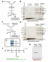
Figure 1. Purification of factors used in this study.
(A) Schematic outlining the purification of human TFIIF and a Coomassie stained gel of the resulting purified material. (B) Schematic outlining the purification of endogenous human pol II and a silver stained gel of the purified complex. * Probable breakdown product of RPB1. This band is recognized by RPB1 antibodies in immunoblot experiments and its presence increases in proportion to the number of times the purified pol II sample is freeze-thawed. Pol II utilized for EM analysis was not freeze-thawed, whereas the silver-stained pol II sample shown here was freeze-thawed once. (C) Purification overview for human VP16-Mediator and a silver stained gel of the purified material. Also shown is a glycerol gradient purification of this sample (right). Mediator-containing fractions (13–15) that were subjected to MS analysis (Table 1) are highlighted.
Figure 2
Figure 2. Purification and activity of Mediator–pol II–TFIIF or the Mediator–pol II binary complex.
(A) Schematic outlining the isolation of the Mediator–pol II–TFIIF assembly from individually purified components. Note Mediator was purified bound to the VP16 activation domain (residues 411–490). (B) Silver stained polyacrylamide gel of glycerol gradient fractions from the purification outlined in (A). Subunits are labeled at right. Fraction A denotes the fraction containing the 1.9 MDa Mediator–pol II–TFIIF assembly. (C) Purification scheme used for isolation of the Mediator–pol II binary complex. (D) Silver stained gel of the odd fractions of the glycerol gradient resulting from the purification outlined in (C). Mediator and pol II subunits are listed at the right. Fraction B denotes the sample containing Mediator–pol II. (E) Western blot analysis of pol II (anti-RPB1), Mediator (anti-MED23), and TFIIF (anti-Rap74), which confirms the presence of TFIIF in fraction A and the absence of TFIIF in fraction B. (F) Schematic of the transcription assay used to investigate the activity of the isolated Mediator–pol II assemblies. Note that Mediator, pol II, and TFIIF are excluded from these assays. (G) The isolated Mediator–pol II–TFIIF assembly is transcriptionally active. In vitro transcription indicates the isolated Mediator–pol II–TFIIF assembly (fraction A) can reconstitute activated transcription from reactions lacking these factors, whereas a Mediator–pol II assembly lacking TFIIF cannot (fraction B).
Figure 3
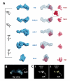
Figure 3. RNA polymerase II adopts a stable orientation within the Mediator–pol II–TFIIF assembly.
(A) Left: different views (rotation shown at left) of the cryo-EM 3D reconstruction of the Mediator–pol II–TFIIF assembly, rendered to 1.8 MDa; center: the pol II crystal structure (red; PDB 1Y1V) is shown docked within the Mediator–pol II–TFIIF cryo-EM map (blue mesh); right: the pol II crystal structure displayed on its own, with characteristic pol II orientations denoted in red font. Note the docked pol II crystal structure shows only the polypeptide backbone and does not correspond to the overall electron density map, whereas the cryo-EM map represents electron density (i.e. a space-filling rendering). A space-filling model of the pol II “front” view is shown (inset) for reference. (B) The top view of the Mediator–pol II–TFIIF assembly cryo-EM structure rendered in the “solid” view using Chimera . Bright areas indicate higher protein density. This rendering allows clear visualization of the pol II cleft. (C) A reprojection view (panel 1) of the Mediator–pol II–TFIIF 3D reconstruction that further highlights the location of the pol II cleft in the structure; panel 2 shows Mediator–pol II–TFIIF in the same orientation, rendered in the “solid” view using Chimera.
Figure 4
Figure 4. Pol II does not stably orient within Mediator in the absence of TFIIF.
The two distinct Mediator–pol II substructures are shown. The location of pol II within each structure—based upon docking the pol II crystal structure (PDB 1Y1V) in Situs —is denoted by the orange sphere. The orientation of pol II, however, could not be reliably determined from docking or projection matching calculations; it appears that multiple pol II orientations exist in the absence of TFIIF.
Figure 5
Figure 5. Structural model of the entire, 3.5 MDa human PIC.
(A) The “front” and “side 1” views of the Mediator–pol II–TFIIF cryo-EM map (blue mesh) are shown. The docked pol II enzyme is shown in red. The locations of TBP, TFIID, TFIIA, TFIIB, TFIIF, TFIIE, and TFIIH are superimposed upon the cryo-EM map and are based upon existing crystallography, EM, and cross-linking studies –,. The likely path of upstream and downstream promoter DNA is also shown (dashed line). (B) The binding site for TFIIS is shown, along with the NTP entry and RNA exit sites within the assembly ,. The putative location of the pol II CTD is shown in green (see text). The Mediator densities labeled “1” and “2” correspond to the head domain. The asterisk denotes the site from which the pol II CTD extends from the enzyme. (C) Ribbon diagram of pol II alone, shown in the same orientation as in (B). Individual pol II subunits colored as shown.
Figure 6
Figure 6. Pol II-induced structural rearrangements block potential Mediator-CDK8 subcomplex interactions.
(A) Two views of the human CDK8 subcomplex . (B) Structure of the human Mediator complex, bound to the activation domain of VP16 . The CDK8 module hook domain binds the leg region (boxed area) of Mediator. Note the structural complementarity between the CDK8 submodule hook domain and the Mediator leg domain. Rearrangements in the leg region that ablate structural complementarity with the CDK8 submodule hook domain occur upon pol II binding in the presence (C) or absence (D,E) of TFIIF.

References

    1. Gilmour D. S. Promoter proximal pausing on genes in metazoans. Chromosoma. 2009;118:1–10. - PubMed
    1. Hahn S. Structure and mechanism of the RNA polymerase II transcription machinery. Nat Struct Mol Biol. 2004;11:394–403. - PMC - PubMed
    1. Baek H. J, Kang Y. K, Roeder R. G. Human Mediator enhances basal transcription by facilitating recruitment of transcription factor IIB during preinitiation complex assembly. J Biol Chem. 2006;281:15172–15181. - PubMed
    1. Cantin G. T, Stevens J. L, Berk A. J. Activation domain-mediator interactions promote transcription preinitiation complex assembly on promoter DNA. Proc Natl Acad Sci U S A. 2003;100:12003–12008. - PMC - PubMed
    1. Naar A. M, Taatjes D. J, Zhai W, Nogales E, Tjian R. Human CRSP interacts with RNA polymerase II CTD and adopts a specific CTD-bound conformation. Genes & Development. 2002;16:1339–1344. - PMC - PubMed

Publication types

MeSH terms