. 2011 Mar;9(3):e1001034.
doi: 10.1371/journal.pbio.1001034. Epub 2011 Mar 29.

A structural and mutagenic blueprint for molecular recognition of strychnine and d-tubocurarine by different cys-loop receptors

Affiliations

A structural and mutagenic blueprint for molecular recognition of strychnine and d-tubocurarine by different cys-loop receptors

Marijke Brams et al. PLoS Biol. 2011 Mar.

Abstract

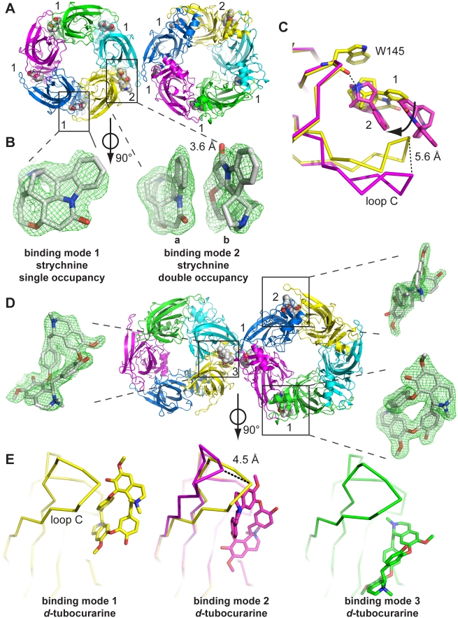
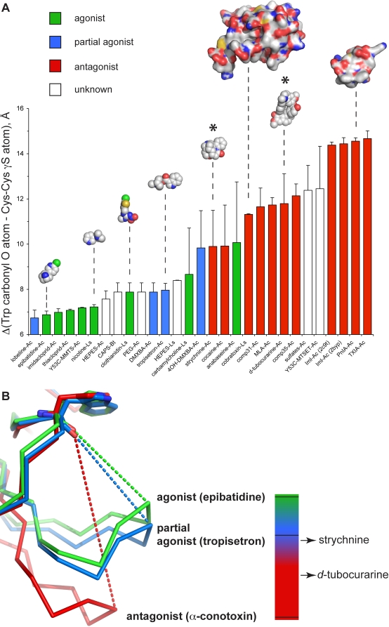
Cys-loop receptors (CLR) are pentameric ligand-gated ion channels that mediate fast excitatory or inhibitory transmission in the nervous system. Strychnine and d-tubocurarine (d-TC) are neurotoxins that have been highly instrumental in decades of research on glycine receptors (GlyR) and nicotinic acetylcholine receptors (nAChR), respectively. In this study we addressed the question how the molecular recognition of strychnine and d-TC occurs with high affinity and yet low specificity towards diverse CLR family members. X-ray crystal structures of the complexes with AChBP, a well-described structural homolog of the extracellular domain of the nAChRs, revealed that strychnine and d-TC adopt multiple occupancies and different ligand orientations, stabilizing the homopentameric protein in an asymmetric state. This introduces a new level of structural diversity in CLRs. Unlike protein and peptide neurotoxins, strychnine and d-TC form a limited number of contacts in the binding pocket of AChBP, offering an explanation for their low selectivity. Based on the ligand interactions observed in strychnine- and d-TC-AChBP complexes we performed alanine-scanning mutagenesis in the binding pocket of the human α1 GlyR and α7 nAChR and showed the functional relevance of these residues in conferring high potency of strychnine and d-TC, respectively. Our results demonstrate that a limited number of ligand interactions in the binding pocket together with an energetic stabilization of the extracellular domain are key to the poor selective recognition of strychnine and d-TC by CLRs as diverse as the GlyR, nAChR, and 5-HT(3)R.


Conflict of interest statement

The authors have declared that no competing interests exist.

Figures

Figure 1
Figure 1. Introduction.
(A) Structure formulas of strychnine and d-tubocurarine (d-TC). (B) Cartoon representation of the acetylcholine binding site in AChBP, which consists of the principal (+) face and complementary (−) face. Different colors are used to highlight loops that form the binding site: loop A (red), B (green), C (yellow), D (magenta), E (blue), and F (orange). Individual residues contributing to loops A–F are indicated according to their numbering in Aplysia AChBP.
Figure 2
Figure 2. X-ray crystal structures of AChBP complexes with strychnine and d-tubocurarine.
(A) Crystal structure of Aplysia AChBP in complex with strychnine as seen along the 5-fold symmetry axis. The asymmetric unit contains 1 pentamer, which interacts with a neighboring pentamer in the crystal packing through an interaction that involves 2 C-loops. At one of these C-loops the ligand binding pocket is occupied by 2 strychnine molecules (indicated with ‘2’). All 4 other binding pockets contain a single strychnine molecule (indicated with ‘1’). (B) Electron density for strychnine molecules. 2Fo−Fc density for single occupancy was contoured at 1.5 sigma, and for double occupancy the sigma level was 0.8. Ligands are shown perpendicular to the 5-fold symmetry axis. (C) Superposition of the 2 binding modes that occur in the AChBP-strychnine complex. The yellow model shows a detailed view of a principal subunit with a single strychnine molecule bound, and the magenta model shows occupancy by two strychnine molecules. Double occupancy results in an outward movement of loop C by 5.6 Å and a rotation of one strychnine molecule around the N-atom involved in the hydrogen bond with the W145 carbonyl. (D) Crystal structure of Aplysia AChBP in complex with d-TC as seen along the 5-fold symmetry axis. The asymmetric unit contains 2 pentamers, which also interact through an interface formed by 2 neighboring C-loops. The 10 ligand binding pockets are characterized by the occupancy of d-TC molecules in 3 different binding orientations. The predominant binding mode is binding mode 1, which is present in most binding pockets, but with varying degree of ligand occupancy. Only sites with full occupancy of binding mode 1 are indicated with ‘1.’ Ligands in binding modes 2 and 3 have full occupancy and occur only once. Electron density for d-TC molecules is shown as 2Fo−Fc density contoured at a sigma level of 1. (E) Comparison of different ligand orientations for d-TC relative to the principal subunits. Binding mode 1 is shown in yellow, mode 2 in magenta, and mode 3 in green. Occupancy of d-TC in binding mode 2 results in a relative outward displacement of loop C by 4.5 Å compared to binding mode 1.
Figure 3
Figure 3. Comparative analysis of C-loop conformation.
(A) For each AChBP crystal structure determined to date we quantified the closure of loop C as the distance between the carbonyl oxygen of the conserved Trp residue of loop A and the sulfur atom of the first cysteine involved in the vicinal disulfide bridge formation (W145 and C188 in Ac-AChBP, W143 and C187 in Ls-AChBP, and W142 and C186 in Bt-AChBP). Bars are colored according to the mode of action of each ligand on nAChRs (agonist, green; partial agonist, blue; antagonist, red; unknown, white). Space fill models are shown for selected ligands. Surface representations were used for α-cobratoxin and α-conotoxin PnIA (A10L, D14K). C-loop conformation for strychnine and d-TC complexes are indicated with *. (B) Superposition of crystal structures of a prototype agonist (epibatidine), partial agonist (tropisetron), and antagonist (α-conotoxin). The dashed lines indicate the distance measure, which is plotted in panel A. The scale bar on the right gives a visually intuitive interpretation of the mode of action for a compound based on C-loop closure. The C-loop contraction for the 2 co-crystal structures reported in this study is indicated with arrows.
Figure 4
Figure 4. Structural recognition of strychnine and d-tubocurarine in AChBP.
(A) Detailed view of the ligand binding pocket for single occupancy by strychnine. Principal face (yellow) and complementary face (blue) are shown in ribbon representation. Amino acids involved in ligand-receptor contacts are shown in sticks. (B) Same as in (A) for double occupancy by strychnine. The same amino acids as in (A) are involved in contacts, except for 3 additional contacts formed by Y186, R57, and T34. (C) Detailed view of the amino acids involved in ligand-receptor interactions with d-TC in binding mode 1. Principal subunit is shown in yellow ribbon and complementary subunit in blue ribbon representation. (D) Detailed view of amino acids forming ligand-receptor contacts for d-TC in binding mode 2. The same residues as in (C) are involved except an additional contact with K141 is formed. Black dashed lines indicate hydrogen bonds. The white dashed line in panel D indicates a polar interaction between the quaternary amine group of d-TC and the carbonyl oxygen of W145.
Figure 5
Figure 5. Correlation between a fast oscillatory movement of AChBP and C-loop closure.
Analysis of the oscillatory frequency (F c) that describes the movement of AChBP during a molecular dynamic simulation for complexes with strychnine, d-TC, and typical agonists and antagonists for the nAChR. Complexes with agonists (nicotine) show a higher oscillation frequency (shown as grey bars) than antagonists (d-TC and PnIA). A good correlation exists with C-loop closure (same data as in Figure 3A, shown as white squares). No data are shown for C-loop closure of the unliganded state because the C-loop was disordered in our X-ray crystal structure of the apo form (pdb code 2w8e).

References

    1. Sine S. M. Molecular dissection of subunit interfaces in the acetylcholine receptor: identification of residues that determine curare selectivity. Proc Natl Acad Sci U S A. 1993;90:9436–9440. - PMC - PubMed
    1. Papineni R. V, Pedersen S. E. Interaction of d-tubocurarine analogs with the mouse nicotinic acetylcholine receptor. Ligand orientation at the binding site. J Biol Chem. 1997;272:24891–24898. - PubMed
    1. Bertrand D, Ballivet M, Rungger D. Activation and blocking of neuronal nicotinic acetylcholine receptor reconstituted in Xenopus oocytes. Proc Natl Acad Sci U S A. 1990;87:1993–1997. - PMC - PubMed
    1. Jonsson M, Gurley D, Dabrowski M, Larsson O, Johnson E. C, et al. Distinct pharmacologic properties of neuromuscular blocking agents on human neuronal nicotinic acetylcholine receptors: a possible explanation for the train-of-four fade. Anesthesiology. 2006;105:521–533. - PubMed
    1. Hope A. G, Belelli D, Mair I. D, Lambert J. J, Peters J. A. Molecular determinants of (+)-tubocurarine binding at recombinant 5-hydroxytryptamine3A receptor subunits. Mol Pharmacol. 1999;55:1037–1043. - PubMed

Publication types

MeSH terms

LinkOut - more resources