. 2011 May;41(5):1399-409.
doi: 10.1002/eji.200939841.

TLR2 signaling improves immunoregulation to prevent type 1 diabetes

Affiliations

TLR2 signaling improves immunoregulation to prevent type 1 diabetes

Christophe M Filippi et al. Eur J Immunol. 2011 May.

Abstract

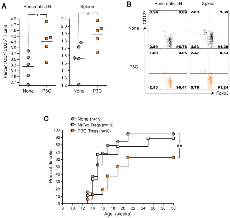
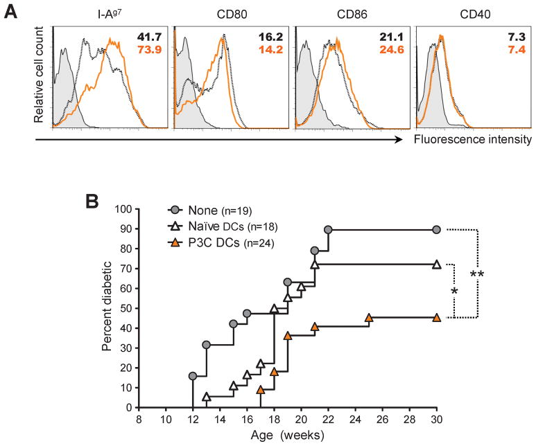
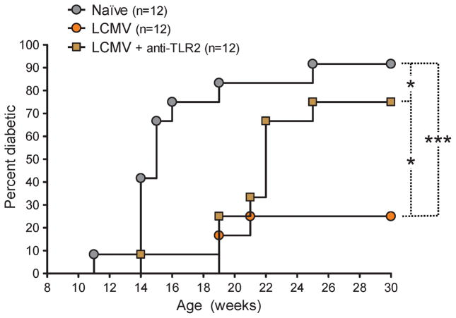
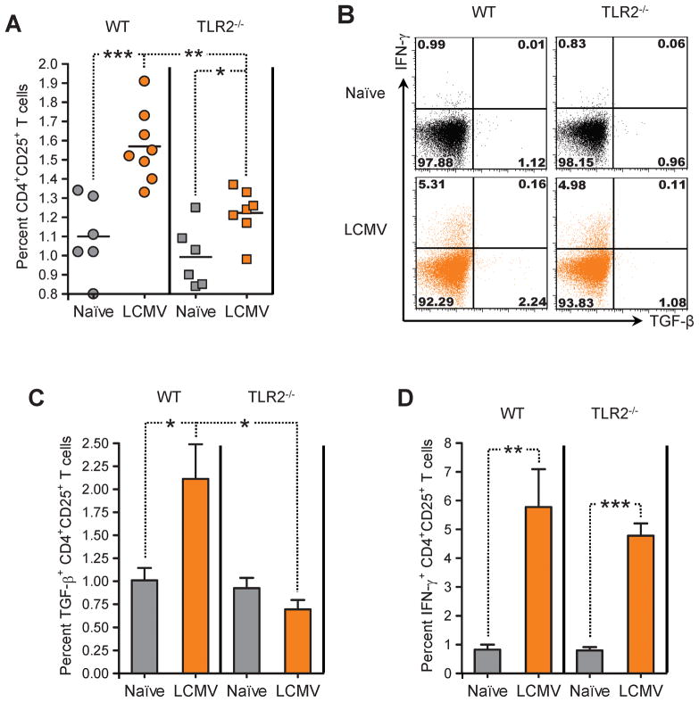
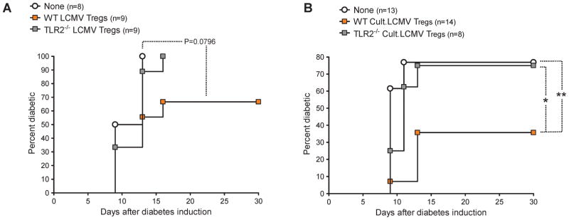
Signaling through TLR2 promotes inflammation and modulates CD4(+) CD25(+) Tregs. We assessed mechanistically how this molecule would alter immunoregulation in type 1 diabetes (T1D). We also asked whether TLR2 may be involved in our recent discovery that viral infection can protect from autoimmune diabetes by expanding and invigorating Tregs. Treatment of prediabetic mice with a synthetic TLR2 agonist diminished T1D and increased the number and function of CD4(+) CD25(+) Tregs, also conferring DCs with tolerogenic properties. TLR2 ligation also promoted the expansion of Tregs upon culture with DCs and ameliorated their capacity to prevent the disease. Protection from T1D by lymphocytic choriomeningitis virus (LCMV) infection depended on TLR2. LCMV increased the frequency of CD4(+) CD25(+) Tregs and their production of TGF-β more significantly in WT than TLR2-deficient mice. Furthermore, LCMV infection in vivo or LCMV-infected DCs in vitro rendered, via TLR2, CD4(+) CD25(+) Tregs capable of diminishing T1D. We identify novel mechanisms by which TLR2 promotes immunoregulation and controls autoimmune diabetes in naïve or infected hosts. This work should help understand T1D etiology and develop novel immune-based therapeutic interventions.


Figures

Figure 1
Figure 1. Activation of TLR2 signaling by P3C in prediabetic NOD mice diminishes type 1 diabetes
(A) Cumulative diabetes incidence over time in NOD mice injected at 9 weeks of age with either saline (None) or 100 μg P3C twice at 5 days interval (P3C). (B) Insulitis scoring of pancreatic islets from Naïve (None) and P3C-treated (P3C) at 11 and 16 weeks of age.
Figure 2
Figure 2. Activation of TLR2 signaling by P3C in prediabetic NOD mice increases the frequency of CD4+CD25+ Tregs and confers them with protective capacity in type 1 diabetes
(A) Percentage of CD4+CD25+ T cells in the pancreatic LN and spleen of individual 12-week-old NOD mice injected 21 days prior with either saline (None) or 100 μg P3C (P3C) twice at 5 days interval, measured by flow cytometry. (B) Representative flow cytometry dot plots of Foxp3 and CD127 expression by CD4+CD25+ T cells in the pancreatic LN and spleen of individual 12-week-old NOD mice left untreated (None) or injected 21 days prior with 100 μg P3C twice at 5 days interval. Quadrants were defined based on isotype control stainings showing under 0.2% positive cells for each parameter analyzed. Numbers indicate the percentage of cells in the corresponding quadrants. (C) Cumulative diabetes incidence over time in NOD mice left untreated (None, gray circles) or injected at 12 weeks of age with 106 CD4+CD25+ Tregs purified from age-matched NOD donors injected at 9 weeks of age with either saline (Naïve Tregs, white squares) or 100 μg P3C twice at 5 days interval (P3C Tregs, orange squares).
Figure 3
Figure 3. Activation of TLR2 signaling by P3C in prediabetic NOD mice confers DCs with protective capacity in type 1 diabetes
(A) Flow cytometry histograms plots of I-Ag7, CD80, CD86, and CD40 expression by CD11c+ cells purified from 9-week-old NOD mice left untreated (black dotted lines) or injected 15 h prior with 100 μg P3C (orange lines). Filled histograms represent isotype control staining. Numbers indicate the MFI of the parameter analyzed. (B) Cumulative diabetes incidence over time in NOD mice left untreated (None, gray circles) or injected at 9 weeks of age with 106 CD11c+ cells purified from age-matched NOD injected 15 h prior with either saline (Naïve DCs, white triangles) or 100 μg P3C (P3C DCs, orange triangles).
Figure 4
Figure 4. CD4+CD25+ Tregs cultured with DCs and P3C efficiently prevent type 1 diabetes in NOD mice
CD4+CD25+ T cells were purified from 9-week-old NOD mice and cultured in the presence of DCs (CD11c+ cells purified from age-matched, syngeneic mice) and 10 U/ml rhIL-2 for 6 days with media alone (Cult.None Tregs) or 2 μg/ml P3C (Cult.P3C Tregs). At the end of the culture, the Tregs were separated from the DCs by negative selection of MHC class-II-expressing cells. (A) Fold expansion of CD4+CD25+ T cells over the 6-day culture period (from duplicate samples). (B) Flow cytometry histograms plots of Foxp3, CD25, CD127, and PD-L1 expression by Tregs purified after culture with DCs in the absence (black dotted lines) or presence (orange lines) of P3C. Filled histograms represent isotype control staining. Numbers indicate the MFI of the parameter analyzed. (C) Cumulative diabetes incidence over time in NOD mice left untreated (None, gray circles) or injected at 10 weeks of age with 106 Tregs cultured with DCs in the absence (Cult.Naïve Tregs, white squares) or presence (Cult.P3C Tregs, orange squares) of P3C.
Figure 5
Figure 5. Activation of TLR2 signaling by LCMV infection in prediabetic NOD mice diminishes type 1 diabetes
Cumulative diabetes incidence over time in NOD mice left untreated (None) or infected at 9 weeks of age with LCMV and left untreated (LCMV) or injected simultaneously and 5 days later with 50 μg of TLR2 blocking mAb (LCMV + anti-TLR2).
Figure 6
Figure 6. LCMV infection of TLR2-deficient B6 mice fails to enhance the frequency of CD4+CD25+ Tregs and their production of TGF-β in the spleen
(A) Percentage of CD4+CD25+ T cells in the spleen of individual age-matched WT (circles) or TLR2−/− (squares) B6 mice left untreated (gray symbols) or infected 21 days prior with LCMV (orange symbols), measured by flow cytometry. (B) Representative flow cytometry dot plots of TGF-β and IFN-γ production by CD4+CD25+ T cells in the spleen of individual age-matched WT or TLR2−/− B6 mice left untreated (Naïve) or infected 21 days prior with LCMV, measured by flow cytometry after polyclonal stimulation. Quadrants were defined based on isotype control stainings showing under 0.2% positive cells for each parameter analyzed. Numbers indicate the percentage of cells in the corresponding quadrants. (C–D) Percentage of TGF-β-(C) and IFN-γ-producing CD4+CD25+ T cells in the spleen of WT or TLR2−/− B6 mice left untreated (Naïve) or infected 21 days prior with LCMV, measured by flow cytometry after polyclonal stimulation (4 to 7 mice per group).
Figure 7
Figure 7. LCMV confers CD4+CD25+ Tregs with protective capacity in type 1 diabetes via TLR2 and DCs
(A) Cumulative diabetes incidence over time in B6 RIP-GP mice infected with LCMV and left untreated (None) or simultaneously injected with 1.5×10⁶ CD4+CD25+ T cells purified from WT (WT LCMV Tregs) or TLR2−/− B6 mice (TLR2−/−LCMV Tregs) infected 21 days prior with LCMV. (B) CD4+CD25+ T cells purified from WT or TLR2−/− B6 mice infected 21 days prior with LCMV were cultured for 6 days with DCs purified from WT or TLR2−/− B6 mice, respectively, infected 48 h prior with LCMV (Cult.LCMV Tregs). Shown is cumulative diabetes incidence over time in B6 RIP-GP mice infected with LCMV and left untreated (None) or simultaneously injected with 5×10⁵ Tregs from WT (WT Cult.LCMV Tregs) or TLR2−/− (TLR2−/− Cult.LCMV Tregs) cultures.

References

    1. Eisenbarth GS. Type I diabetes mellitus. A chronic autoimmune disease. N Engl J Med. 1986;314:1360–1368. - PubMed
    1. Filippi CM, von Herrath MG. Viral trigger for type 1 diabetes: pros and cons. Diabetes. 2008;57:2863–2871. - PMC - PubMed
    1. Foulis AK, Jackson R, Farquharson MA. The pancreas in idiopathic Addison’s disease--a search for a prediabetic pancreas. Histopathology. 1988;12:481–490. - PubMed
    1. Horwitz MS, Ilic A, Fine C, Balasa B, Sarvetnick N. Coxsackieviral-mediated diabetes: induction requires antigen-presenting cells and is accompanied by phagocytosis of beta cells. Clin Immunol. 2004;110:134–144. - PubMed
    1. Oldstone MB, Nerenberg M, Southern P, Price J, Lewicki H. Virus infection triggers insulin-dependent diabetes mellitus in a transgenic model: role of anti-self (virus) immune response. Cell. 1991;65:319–331. - PubMed

Publication types

MeSH terms