

Cephalometric measurements from 3D reconstructed images compared with conventional 2D images
- PMID: 21469969
- PMCID: PMC8916191
- DOI: 10.2319/121210-717.1
Cephalometric measurements from 3D reconstructed images compared with conventional 2D images
Abstract
Objective: To assess whether the values of different measurements taken on three-dimensional (3D) reconstructions from cone-beam computed tomography (CBCT) are comparable with those taken on two-dimensional (2D) images from conventional lateral cephalometric radiographs (LCRs) and to examine if there are differences between the different types of CBCT software when taking those measurements.
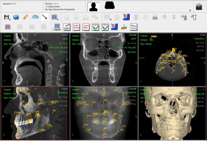
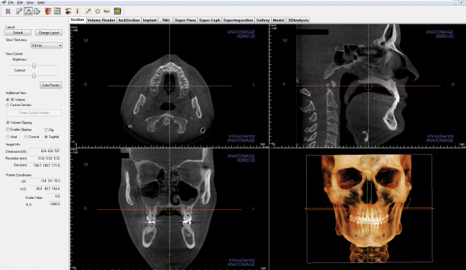
Material and methods: Eight patients were selected who had both an LRC and a CBCT. The 3D reconstructions of each patient in the CBCT were evaluated using two different software packages, NemoCeph 3D and InVivo5. An observer took 10 angular and 3 linear measurements on each of the three types of record on two different occasions.
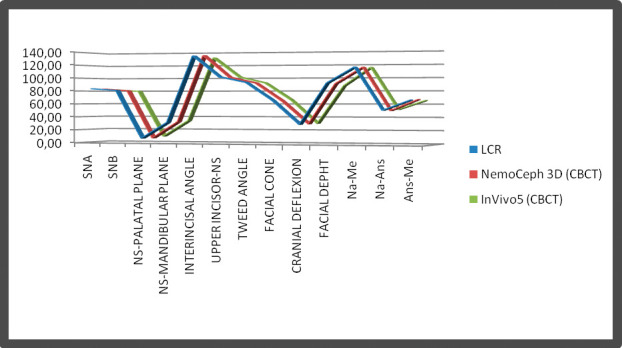
Results: Intraobserver reliability was high except for the mandibular plane and facial cone (from the LCR), the Na-Ans distance (using NemoCeph 3D), and facial cone and the Ans-Me distance (using InVivo5). No statistically significant differences were found for the angular and linear measurements between the LCRs and the CBCTs for any measurement, and the correlation levels were high for all measurements.
Conclusion: No statistically significant differences were found between the angular and linear measurements taken with the LCR and those taken with the CBCT. Neither were there any statistically significant differences between the angular or linear measurements using the two CBCT software packages.
Figures
References
-
- Arai Y, Tammisalo E, Iwai K, Hashimoto K, Shinoda K. Development of a compact computed tomographic apparatus for dental use. Dentomaxillofac Radiol. 1999;28:245–248. - PubMed
-
- Mozzo P, Procacci C, Tacconi A, Martini P. T, Andreis I. A. A new volumetric CT machine for dental imaging based on the cone-beam technique: preliminary results. Eur Radiol. 1998;8:1558–1564. - PubMed
-
- Mah J, Hatcher D. Diagnóstico por imagen craneofacial en ortodoncia. Capítulo 2. In: Grabber T. M, Vanarsdall R. L, Vig K. W. L, editors. Orthodontics Current Principles and Techniques. St Louis, MO: Elsevier; 2005. pp. 71–100.
-
- Kitaura H, Yonetsu K, Kitamori H, Kobayashi K, Nakamura T. Standardization of 3-D CT measurements for length and angles by matrix transformation in the 3-D coordinate system. Cleft Palate Craniofac J. 2000;37:349–356. - PubMed
-
- Pinsky H. M, Dyda S, Pinsky R. W, Misch K. A, Sarment D. P. Accuracy of three-dimensional measurements using cone-beam CT. Dentomaxillofac Radiol. 2006;35:410–416. - PubMed
Publication types
MeSH terms
LinkOut - more resources
Full Text Sources
