

Differential analysis of glioblastoma multiforme proteome by a 2D-DIGE approach
- PMID: 21470419
- PMCID: PMC3083325
- DOI: 10.1186/1477-5956-9-16
Differential analysis of glioblastoma multiforme proteome by a 2D-DIGE approach
Abstract
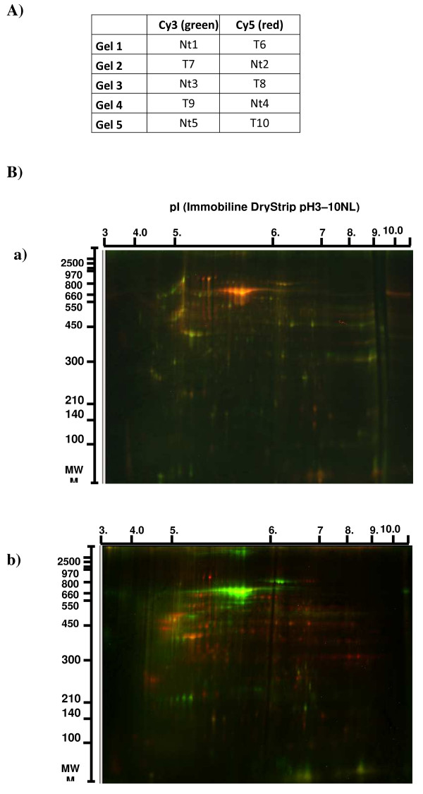
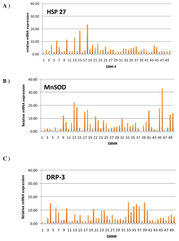
Background: Genomics, transcriptomics and proteomics of glioblastoma multiforme (GBM) have recently emerged as possible tools to discover therapeutic targets and biomarkers for new therapies including immunotherapy. It is well known that macroscopically complete surgical excision, radiotherapy and chemotherapy have therapeutic limitations to improve survival in these patients. In this study, we used a differential proteomic-based technique (2D-Difference Gel Electrophoresis) coupled with matrix-assisted laser desorption/ionization-time of flight (MALDI-TOF) mass spectrometry to identify proteins that may serve as brain tumor antigens in new therapeutic assays. Five samples of patients presenting a GBM and five samples of microscopically normal brain tissues derived from brain epileptic surgery specimen were labeled and run in 2D-PAGE (Two-Dimensional Polyacrylamide Gel Electrophoresis) with an internal pool sample on each gel. Five gels were matched and compared with DIA (Difference In-gel Analysis) software. Differential spots were picked, in-gel digested and peptide mass fingerprints were obtained.
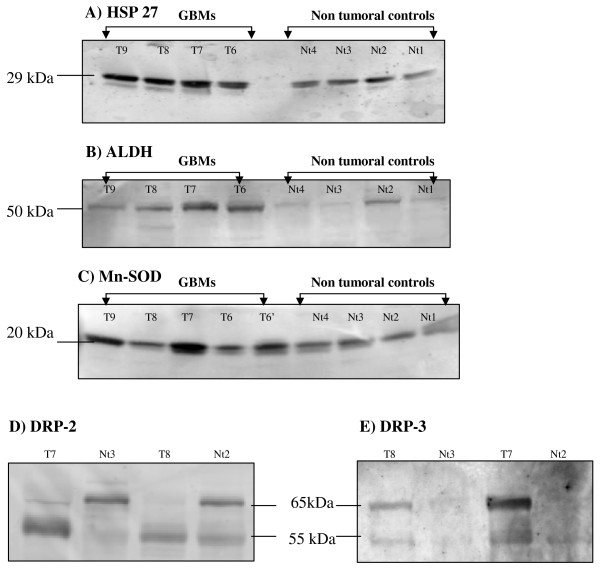
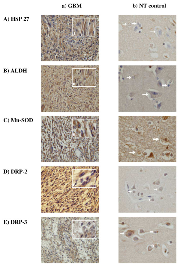
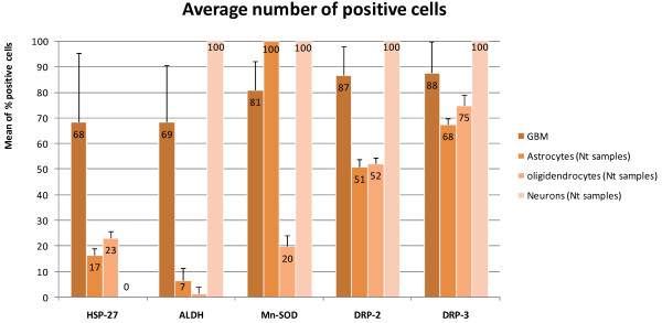
Results: From 51 protein-spots significantly up-regulated in GBM samples, mass spectrometry (MS) identified twenty-two proteins. The differential expression of a selected protein set was first validated by western-blotting, then tested on large cohorts of GBM specimens and non-tumor tissues, using immunohistochemistry and real-time RT-PCR.
Conclusions: Our results confirmed the importance of previously described proteins in glioma pathology and their potential usefulness as biological markers but also revealed some new interesting targets for future therapies.
Figures
Comment in
- Biomark Med. 2011 Aug;5(4):423-5
Similar articles
-
Investigation of serum proteome alterations in human glioblastoma multiforme.Proteomics. 2012 Aug;12(14):2378-90. doi: 10.1002/pmic.201200002. Proteomics. 2012. PMID: 22684992
-
Identification of differentially-expressed proteins between early submucosal non-invasive and invasive colorectal cancer using 2D-DIGE and mass spectrometry.Int J Immunopathol Pharmacol. 2011 Oct-Dec;24(4):849-59. doi: 10.1177/039463201102400404. Int J Immunopathol Pharmacol. 2011. PMID: 22230392
-
Proteomics identifies differentially expressed proteins in neonatal murine thymus compared with adults.Proteome Sci. 2012 Nov 8;10(1):65. doi: 10.1186/1477-5956-10-65. Proteome Sci. 2012. PMID: 23134655 Free PMC article.
-
Proteome analysis of human liver tumor tissue by two-dimensional gel electrophoresis and matrix assisted laser desorption/ionization-mass spectrometry for identification of disease-related proteins.Electrophoresis. 2002 Dec;23(24):4142-56. doi: 10.1002/elps.200290032. Electrophoresis. 2002. PMID: 12481271
-
Two-Dimensional Gel Electrophoresis and 2D-DIGE.Methods Mol Biol. 2018;1664:3-14. doi: 10.1007/978-1-4939-7268-5_1. Methods Mol Biol. 2018. PMID: 29019120 Review.
Cited by
-
Cluster and Principal Component Analysis of Human Glioblastoma Multiforme (GBM) Tumor Proteome.Iran J Cancer Prev. 2014 Spring;7(2):87-95. Iran J Cancer Prev. 2014. PMID: 25250155 Free PMC article.
-
Identification of Ubiquitination-Associated Proteins Using 2D-DIGE.Methods Mol Biol. 2023;2596:83-96. doi: 10.1007/978-1-0716-2831-7_6. Methods Mol Biol. 2023. PMID: 36378432
-
Proteins inform survival-based differences in patients with glioblastoma.Neurooncol Adv. 2020 Mar 17;2(1):vdaa039. doi: 10.1093/noajnl/vdaa039. eCollection 2020 Jan-Dec. Neurooncol Adv. 2020. PMID: 32642694 Free PMC article.
-
The Study of "Dihydropyrimidinase Related Proteins (DRPs)" Expression Changes Influence in Malignant Astrocytoma Brain Tumor.Iran J Cancer Prev. 2014 Summer;7(3):130-6. Iran J Cancer Prev. 2014. PMID: 25250163 Free PMC article.
-
PLEK2, RRM2, GCSH: A Novel WWOX-Dependent Biomarker Triad of Glioblastoma at the Crossroads of Cytoskeleton Reorganization and Metabolism Alterations.Cancers (Basel). 2021 Jun 12;13(12):2955. doi: 10.3390/cancers13122955. Cancers (Basel). 2021. PMID: 34204789 Free PMC article.
References
LinkOut - more resources
Full Text Sources
