

Implications of cultured periodontal ligament cells for the clinical and experimental setting: a review
- PMID: 21470594
- PMCID: PMC3132241
- DOI: 10.1016/j.archoralbio.2011.03.003
Implications of cultured periodontal ligament cells for the clinical and experimental setting: a review
Abstract
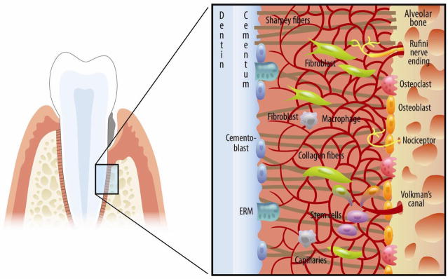
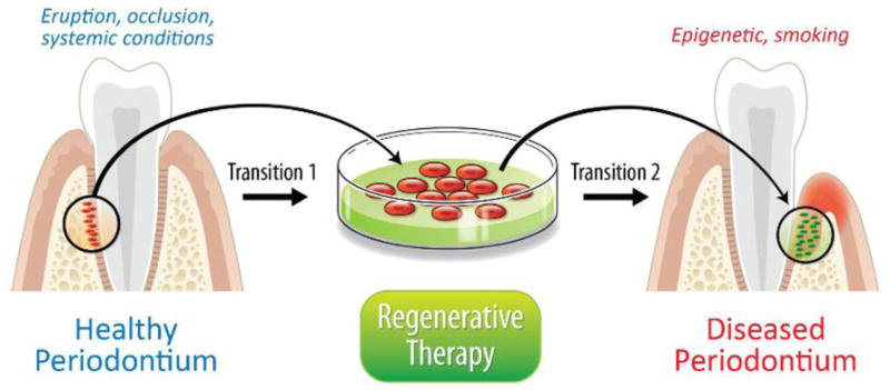
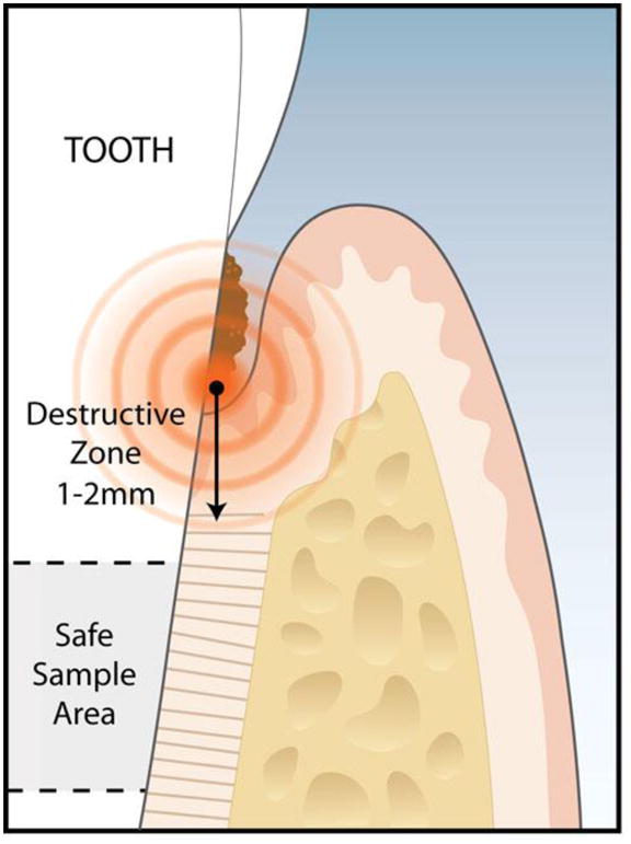
The periodontal ligament (PDL) is a key contributor to the process of regeneration of the periodontium. The heterogeneous nature of the PDL tissue, its development during early adulthood, and the different conditions to which the PDL tissue is exposed to in vivo impart on the PDL unique characteristics that may be of consequence during its cultivation in vitro. Several factors affecting the in vivo setting influence the behaviour of PDL fibroblasts in culture. The purpose of this review is to address distinct factors that influence the behaviour of PDL fibroblasts in culture -in vivo-in vitro transitions, cell identification/isolation markers, primary PDL cultures and cell lines, tooth-specific factors, and donor-specific factors. Based on the reviewed studies, the authors recommendations include the use of several identification markers to confirm cell identity, use of primary cultures at early passage to maintain unique PDL heterogeneic characteristics, and noting donor conditions such as age, systemic health status, and tooth health status. Continued efforts will expand our understanding of the in vitro and in vivo behaviour of cells, with the goal of orchestrating optimal periodontal regeneration. This understanding will lead to improved evidence-based rationales for more individualized and predictable periodontal regenerative therapies.
Copyright © 2011 Elsevier Ltd. All rights reserved.
Figures
Similar articles
-
Development and characterization of a transformed human periodontal ligament cell line.J Periodontol. 1997 Nov;68(11):1054-62. doi: 10.1902/jop.1997.68.11.1054. J Periodontol. 1997. PMID: 9407397
-
Comparison of characteristics of periodontal ligament cells obtained from outgrowth and enzyme-digested culture methods.Arch Oral Biol. 2011 Apr;56(4):380-8. doi: 10.1016/j.archoralbio.2010.10.013. Epub 2010 Dec 8. Arch Oral Biol. 2011. PMID: 21144495
-
Flow cytometry analysis of gingival and periodontal ligament cells.J Dent Res. 1998 Apr;77(4):555-64. doi: 10.1177/00220345980770040801. J Dent Res. 1998. PMID: 9539458
-
Dental stem cell dynamics in periodontal ligament regeneration: from mechanism to application.Stem Cell Res Ther. 2024 Oct 31;15(1):389. doi: 10.1186/s13287-024-04003-9. Stem Cell Res Ther. 2024. PMID: 39482701 Free PMC article. Review.
-
Physiological features of periodontal regeneration and approaches for periodontal tissue engineering utilizing periodontal ligament cells.J Biosci Bioeng. 2007 Jan;103(1):1-6. doi: 10.1263/jbb.103.1. J Biosci Bioeng. 2007. PMID: 17298893 Review.
Cited by
-
Induction of IL-6 and MMP-8 in human periodontal fibroblasts by static tensile strain.Clin Oral Investig. 2014 Apr;18(3):901-8. doi: 10.1007/s00784-013-1032-1. Epub 2013 Jul 14. Clin Oral Investig. 2014. PMID: 23851938
-
Central role of pyrophosphate in acellular cementum formation.PLoS One. 2012;7(6):e38393. doi: 10.1371/journal.pone.0038393. Epub 2012 Jun 4. PLoS One. 2012. PMID: 22675556 Free PMC article.
-
Gene Expression Profile in Immortalized Human Periodontal Ligament Fibroblasts Through hTERT Ectopic Expression: Transcriptome and Bioinformatic Analysis.Front Mol Biosci. 2021 Jun 1;8:679548. doi: 10.3389/fmolb.2021.679548. eCollection 2021. Front Mol Biosci. 2021. PMID: 34141725 Free PMC article.
-
Phytic acid effect on periodontal ligament fibroblast: An in-vitro study.PLoS One. 2023 Dec 14;18(12):e0295612. doi: 10.1371/journal.pone.0295612. eCollection 2023. PLoS One. 2023. PMID: 38096253 Free PMC article.
-
Establishing a technique for isolation and characterization of human periodontal ligament derived mesenchymal stem cells.Saudi Dent J. 2021 Nov;33(7):693-701. doi: 10.1016/j.sdentj.2020.04.007. Epub 2020 Apr 19. Saudi Dent J. 2021. PMID: 34803321 Free PMC article.
References
-
- Wang HL, Greenwell H, Fiorellini J, Giannobile W, Offenbacher S, Salkin L, et al. Periodontal regeneration. J Periodontol. 2005 Sep;76(9):1601–22. - PubMed
-
- Bosshardt DD, Sculean A. Does periodontal tissue regeneration really work? Periodontol 2000. 2009;51:208–19. - PubMed
-
- Flores MG, Yashiro R, Washio K, Yamato M, Okano T, Ishikawa I. Periodontal ligament cell sheet promotes periodontal regeneration in athymic rats. J Clin Periodontol. 2008 Dec;35(12):1066–72. - PubMed
-
- Nanci A, Bosshardt DD. Structure of periodontal tissues in health and disease. Periodontol 2000. 2006;40:11–28. - PubMed
-
- Reeve CM, Wentz FM. The prevalence, morphology, and distribution of epithelial rests in the human periodontal ligament. Oral Surg Oral Med Oral Pathol. 1962 Jul;15:785–93. - PubMed
Publication types
MeSH terms
Substances
Grants and funding
LinkOut - more resources
Full Text Sources
