. 2011 Jun;69(6):e50-7.
doi: 10.1016/j.joms.2010.12.049. Epub 2011 Apr 5.

Osteochondral interface regeneration of rabbit mandibular condyle with bioactive signal gradients

Affiliations

Osteochondral interface regeneration of rabbit mandibular condyle with bioactive signal gradients

Nathan H Dormer et al. J Oral Maxillofac Surg. 2011 Jun.

Abstract

Purpose: Tissue engineering solutions focused on the temporomandibular joint (TMJ) have expanded in number and variety during the past decade to address the treatment of TMJ disorders. The existing data on approaches for healing small defects in the TMJ condylar cartilage and subchondral bone, however, are sparse. The purpose of the present study was thus to evaluate the performance of a novel gradient-based scaffolding approach to regenerate osteochondral defects in the rabbit mandibular condyle.

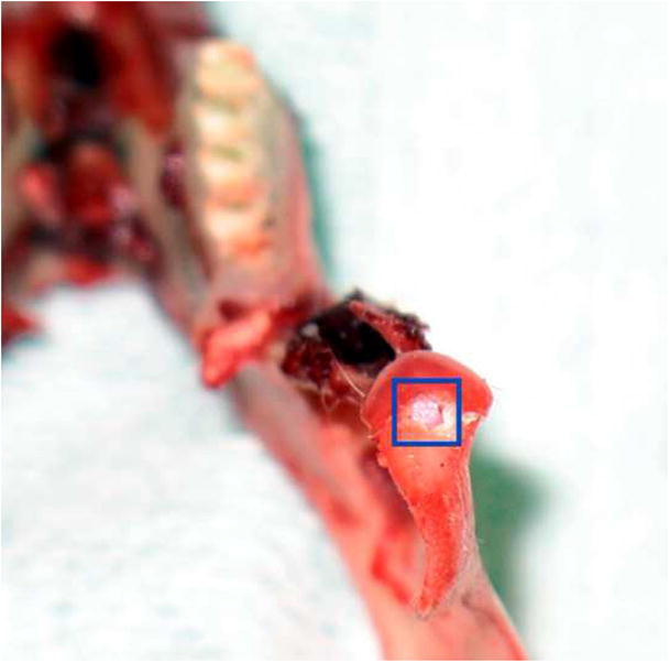
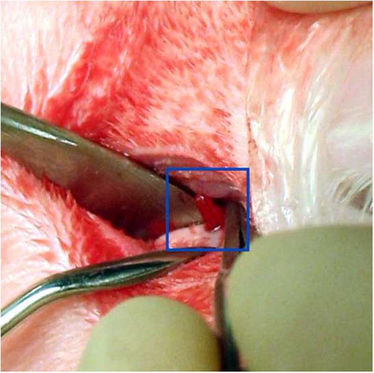
Materials and methods: Miniature bioactive plugs for regeneration of small mandibular condylar defects in New Zealand white rabbits were fabricated. The plugs were constructed from poly(D,L-lactic-co-glycolic acid) microspheres with a gradient transition between cartilage-promoting and bone-promoting growth factors.

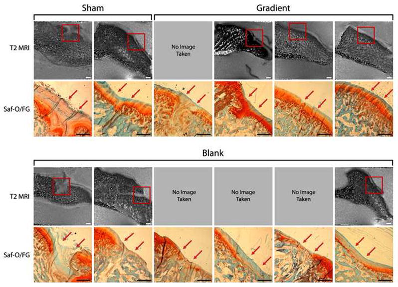
Results: At 6 weeks of healing, the results suggested that the implants provided support for the neosynthesized tissue as evidenced by the histologic and 9.4 T magnetic resonance imaging findings.

Conclusion: The inclusion of bioactive factors in a gradient-based scaffolding design is a promising new treatment strategy for focal defect repair in the TMJ.


Figures

Figure 1
Figure 1
Microsphere and scaffold fabrication process. (A) Microspheres were made from a polymer stream (20% w/v PLGA in DCM) and annular carrier stream (0.5% w/v PVA in DI H2O) with an ultrasonic transducer; (B) Programmable pumps created a gradient in microsphere types; (C) Experimental groups for each time point (six rabbits at twelve weeks). Sham surgeries were performed with no implant, blank groups contained microspheres with no growth factor, gradient groups contained a gradient of BMP-2 and TGF-β1; (D) Microscope image of microsphere-based scaffold with particles of 70 μm. Scale bar = 500 μm.
Figure 1
Figure 1
Microsphere and scaffold fabrication process. (A) Microspheres were made from a polymer stream (20% w/v PLGA in DCM) and annular carrier stream (0.5% w/v PVA in DI H2O) with an ultrasonic transducer; (B) Programmable pumps created a gradient in microsphere types; (C) Experimental groups for each time point (six rabbits at twelve weeks). Sham surgeries were performed with no implant, blank groups contained microspheres with no growth factor, gradient groups contained a gradient of BMP-2 and TGF-β1; (D) Microscope image of microsphere-based scaffold with particles of 70 μm. Scale bar = 500 μm.
Figure 2
Figure 2
Implant placement. (A) The defect was created in the anterior region of the TMJ condyle, as seen in this excised mandible specimen; (B) Implant being press fitted into articulating surface of TMJ condyle..
Figure 2
Figure 2
Implant placement. (A) The defect was created in the anterior region of the TMJ condyle, as seen in this excised mandible specimen; (B) Implant being press fitted into articulating surface of TMJ condyle..
Figure 3
Figure 3
Cartilage Evaluation - T2 MRI images and histological staining of rabbit TMJs at six weeks of time for sham, blank, gradient groups. “SF” denotes Safranin-O/Fast Green for GAGs. Sham and gradient samples earned similar histological scores for cartilage thickness width over defects, whereas blank samples had noticeably thinner neo-cartilage compared to gradient samples, although this was not statistically significant (p = 0.156). Red arrows mark the approximate edges of the original defect. Scale bar = 1.0 mm in MRI and histological images.
Figure 4
Figure 4
Bone Evaluation - histological staining of rabbit TMJs at six weeks for sham, blank, and gradient groups. “AR” denotes Alizarin Red for calcium ions, and “VK” denotes von Kossa for calcium phosphate. There was a slight tendency for subchondral trabeculae in gradient samples to be thicker. Samples are arranged in the exact same order as seen in Figure 3. Red arrows mark the approximate edges of the original defect. Scale bar = 1.0 mm.

References

    1. Hollister SJ, Levy RA, Chu TM, et al. An image-based approach for designing and manufacturing craniofacial scaffolds. Int J Oral Maxillofac Surg. 2000;29:67. - PubMed
    1. Schek RM, Taboas JM, Segvich SJ, et al. Engineered osteochondral grafts using biphasic composite solid free-form fabricated scaffolds. Tissue Eng. 2004;10:1376. - PubMed
    1. Schek RM, Taboas JM, Hollister SJ, Krebsbach PH. Tissue engineering osteochondral implants for temporomandibular joint repair. Orthod Craniofac Res. 2005;8:313. - PubMed
    1. Williams JM, Adewunmi A, Schek RM, et al. Bone tissue engineering using polycaprolactone scaffolds fabricated via selective laser sintering. Biomaterials. 2005;26:4817. - PubMed
    1. Hollister SJ, Lin CY, Saito E, et al. Engineering craniofacial scaffolds. Orthod Craniofac Res. 2005;8:162. - PubMed

Publication types

Substances