. 2011 Jun 1;22(11):1810-23.
doi: 10.1091/mbc.E11-01-0019. Epub 2011 Apr 6.

Glucosidase II and N-glycan mannose content regulate the half-lives of monoglucosylated species in vivo

Affiliations

Glucosidase II and N-glycan mannose content regulate the half-lives of monoglucosylated species in vivo

Ivan D Stigliano et al. Mol Biol Cell. .

Abstract

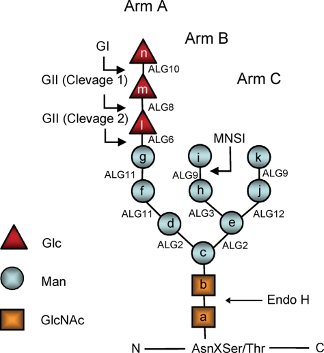
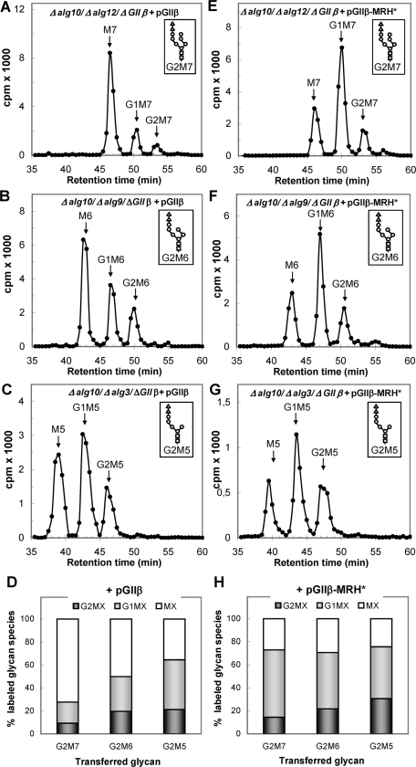
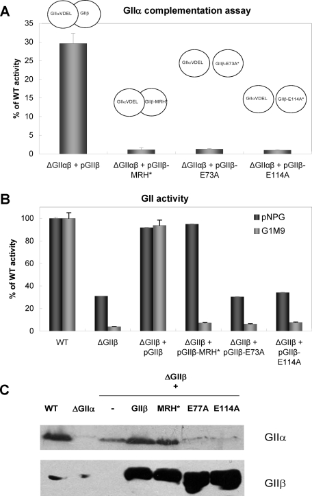
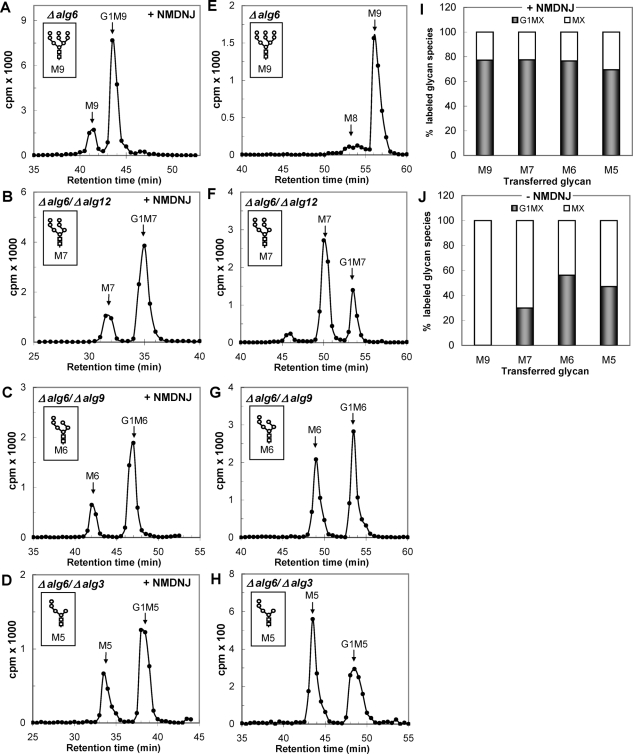
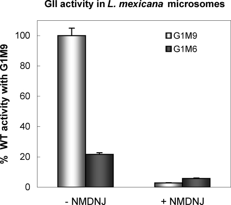
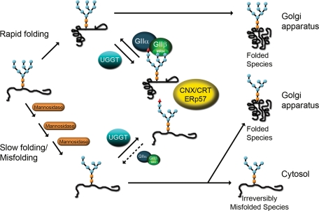
Glucosidase II (GII) sequentially removes the two innermost glucose residues from the glycan (Glc(3)Man(9)GlcNAc(2)) transferred to proteins. GII also participates in cycles involving the lectin/chaperones calnexin (CNX) and calreticulin (CRT) as it removes the single glucose unit added to folding intermediates and misfolded glycoproteins by the UDP-Glc:glycoprotein glucosyltransferase (UGGT). GII is a heterodimer in which the α subunit (GIIα) bears the active site, and the β subunit (GIIβ) modulates GIIα activity through its C-terminal mannose 6-phosphate receptor homologous (MRH) domain. Here we report that, as already described in cell-free assays, in live Schizosaccharomyces pombe cells a decrease in the number of mannoses in the glycan results in decreased GII activity. Contrary to previously reported cell-free experiments, however, no such effect was observed in vivo for UGGT. We propose that endoplasmic reticulum α-mannosidase-mediated N-glycan demannosylation of misfolded/slow-folding glycoproteins may favor their interaction with the lectin/chaperone CNX present in S. pombe by prolonging the half-lives of the monoglucosylated glycans (S. pombe lacks CRT). Moreover, we show that even N-glycans bearing five mannoses may interact in vivo with the GIIβ MRH domain and that the N-terminal GIIβ G2B domain is involved in the GIIα-GIIβ interaction. Finally, we report that protists that transfer glycans with low mannose content to proteins have nevertheless conserved the possibility of displaying relatively long-lived monoglucosylated glycans by expressing GIIβ MRH domains with a higher specificity for glycans with high mannose content.


Figures

FIGURE 1:
FIGURE 1:
Glycan structures. The structure depicted is that of the full-length glycan transferred to Asn residues in N‑glycosylation. Lettering (a, b, c…) follows the order of addition of the monosaccharides in the synthesis of the Dol-P-P derivatives. Alg genes involved in the synthetic process are indicated. GI removes residue n, and GII removes residues m and l. UGGT re-adds residue l. Glycans have the following structures: M9, residues ak; M7, ai; M6, ah; and M5, ag. The respective mono-, di-, and triglucosylated derivatives have, additionally, residues l, l–m, and ln. Because glycans with nine, seven, or six mannoses were released with Endo H, they lack residue a when separated by paper chromatography or HPLC. In contrast, glycans with five mannoses were released with N-glycanase and therefore have residue a. Arm A comprises residues d, f, g, and l–n, arm B comprises residues h and I, and arm C comprises residues j and k.
FIGURE 2:
FIGURE 2:
Glycan patterns synthesized by mutants transferring diglucosylated glycans containing nine to five mannoses. (A) G2M9 (Δalg10); (B) G2M7 (Δalg10/Δalg12); (C) G2M6 (Δalg10/Δalg9), and (D) G2M5 (Δalg10/Δalg3). The structures of the glycans transferred by each mutant are indicated in the corresponding panels. (E) Quantification of the relative amounts of the di-, mono-, and unglucosylated species from panels A–D. In panel A, the label in M8 was added to that in M9 to account for unglucosylated species.
FIGURE 3:
FIGURE 3:
ER GIIα content in S. pombe cells expressing wild-type, mutant, or no GIIβ. Each lane was loaded with 250 μg of microsomal proteins of ΔGIIβ cells or ΔGIIβ cells expressing exogenous GIIβ or GIIβ-MRH* (MRH*). The membrane was blotted using mouse polyclonal anti-GIIα subunit (1:500) and rabbit polyclonal anti-CNX (1:100,000) primary antibodies. Goat HRP anti–mouse or –rabbit IgG (1:5000 and 1:30,000, respectively) were used as secondary antibodies. Reactions were detected by chemiluminescence.
FIGURE 4:
FIGURE 4:
Glycan patterns synthesized by mutants transferring diglucosylated glycans containing nine to five mannoses and lacking GIIβ. (A) G2M9 (Δalg10/ΔGIIβ); (B) G2M7 (Δalg10/Δalg12/ΔGIIβ); (C) G2M6 (Δalg10/Δalg9/ΔGIIβ); (D) G2M5 (Δalg10/Δalg3/ΔGIIβ). The structures of the glycans transferred by each mutant are indicated in the corresponding panels. (E) Quantification of the relative amounts of the di-, mono-, and unglucosylated species from panels A–D.
FIGURE 5:
FIGURE 5:
Glycan patterns synthesized by mutants transferring diglucosylated glycans containing seven to five mannoses and lacking GIIβ but expressing exogenous wild-type GIIβ (A–D) or exogenous GIIβ with a mutated MRH domain (E–H). (A) G2M7 (Δalg10/Δalg12/ΔGIIβ + pGIIβ); (B) G2M6 (Δalg10/Δalg9/ΔGIIβ + pGIIβ); (C) G2M5 (Δalg10/Δalg3/ΔGIIβ + pGIIβ); (E) G2M7 (Δalg10/Δalg12/ΔGIIβ + pGIIβ-MRH*); (F) G2M6 (Δalg10/Δalg9/ΔGIIβ + pGIIβ-MRH*); (G) G2M5 (Δalg10/Δalg3/ΔGIIβ + pGIIβ-MRH*). The structures of the glycans transferred by each mutant are indicated in the corresponding panels. (D and H) Quantification of the relative amounts of the glycans shown in panels A–C (D) and E–G (H).
FIGURE 6:
FIGURE 6:
Mutations in the GIIβ G2B domain affect the GIIα–GIIβ interaction. (A) Complementation assay between GIIα and GIIβ. Microsomes from ΔGIIαβ S. pombe cells transformed with pGIIαVDEL (the source of the GIIα subunit) were preincubated in the presence of 1% Triton X-100 with microsomes from ΔGIIαβ cells expressing wild-type GIIβ (pGIIβ), GIIβ with a mutated MRH domain (pGIIβ-MRH*), or mutated G2B domain (pGIIβ-E73A or pGIIβ-E114A) (sources of the GIIβ subunit). GII activity assays with [14C-Glc]G1M9 were performed as described in Materials and Methods. (B) GII activity in microsomal fractions of S. pombe wild-type, ΔGIIβ mutant cells or the same mutants expressing exogenous wild-type GIIβ or GIIβ with mutated MRH or G2B domains. Assays were performed with pNPG or G1M9 as substrates. In both cases, the activity was stated relative to that of the wild-type strain (100%). (C) Immunodetection of GIIα and GIIβ in microsomal fractions of S. pombe. Each lane was loaded with 250 μg of microsomal proteins from wild type, ΔGIIα or ΔGIIβ transformed with vector alone (–), wild-type GIIβ (GIIβ), or GIIβ bearing mutations in the MRH (MRH*) or G2B domains (E73A and E113A). The membrane was blotted using mouse polyclonal anti-GIIα subunit (1:500) or -GIIβ subunit (1:1000). Goat HRP anti-mouse 1:5000 was used as the secondary antibody. Reactions were detected by chemiluminescence.
FIGURE 7:
FIGURE 7:
Glycan patterns synthesized by mutants transferring unglucosylated glycans containing nine to five mannoses with inhibition of deglucosylation (plus 5 mM NMDNJ) (A–D) or without inhibition of deglucosylation (minus NMDNJ) (E–H). (A and E) M9 (Δalg6); (B and F) M7 (Δalg6/Δalg12); (C and G) M6 (Δalg6/Δalg9); (D and H) M5 (Δalg6/Δalg3). The structures of the glycans transferred by each mutant are indicated in the corresponding panels. (I and J) Quantification of the relative amounts of the glucosylated and nonglucosylated labeled glycans from panels A–D (I) and E–H (J). In panel E, the label in M8 was added to that of M9. The label in the unidentified peak from panel F was omitted for quantification.
FIGURE 8:
FIGURE 8:
Glucose release from either G1M9 or G1M6 by L. mexicana GII. In all cases, the incubation mixtures contained 1 mM 1-deoxymannojirimycin and, where indicated, the same concentration of NMDNJ. The percentage of the total glucose liberated was calculated from each glycan [14C]glucose content (see Materials and Methods). The value for G1M9 was taken as 100%.
FIGURE 9:
FIGURE 9:
Model proposed for GII as an in vivo regulator of misfolded/slow-folding glycoprotein ER permanence. Misfolded/slow-folding species are characterized by an ER mannosidase(s)-catalyzed N-glycan demannosylation. A decrease in N-glycan mannose content significantly diminishes in vivo GII-mediated deglucosylation rates but does not affect in vivo UGGT-mediated glucosylation, thus increasing the possibility of displaying monoglucosylated structures able to interact with CNX/CRT for longer time periods. The exit of irreversibly misfolded glycoproteins from futile CNX cycles most likely will occur, at least in mammalian cells, upon removal of mannose unit g in Arm A (see Figure 1).

Similar articles

Cited by

References

    1. Alfa C, Fantes P, Hyams J, McLeod M, Wabrik E. Experiments with Fission Yeast: A Laboratory Manual. Cold Spring Harbor, NY: Cold Spring Harbor Laboratory Press; 1993.
    1. Arendt CW, Ostergaard HL. Two distinct domains of the β subunit of glucosidase II interact with the catalytic α-subunit. Glycobiology. 2000;10:487–492. - PubMed
    1. Bähler J, Wu JQ, Longtine MS, Shah NG, McKenzie A 3rd, Steever AB, Wach A, Philippsen P, Pringle JR. Heterologous modules for efficient and versatile PCR-based gene targeting in Schizosaccharomyces pombe. Yeast. 1998;14:943–951. - PubMed
    1. Cabral CM, Liu Y, Sifers RN. Dissecting glycoprotein quality control in the secretory pathway. Trends Biochem Sci. 2001;26:619–624. - PubMed
    1. Caramelo JJ, Parodi AJ. How sugars convey information on protein conformation in the endoplasmic reticulum. Sem Cell Dev Biol. 2007;18:10221–10225. - PMC - PubMed

Publication types

MeSH terms