. 2011 Sep;17(9):577-86.
doi: 10.1093/molehr/gar022. Epub 2011 Apr 5.

Ex vivo functional responses to HLA-G differ between blood and decidual NK cells

Affiliations

Ex vivo functional responses to HLA-G differ between blood and decidual NK cells

Richard Apps et al. Mol Hum Reprod. 2011 Sep.

Abstract

Restricted expression of human leucocyte antigen-G (HLA-G) to fetal extravillous trophoblast cells, which invade the decidua during implantation, suggests a role for HLA-G in placentation. In this study, we have investigated several aspects of HLA-G expression and function. Surface levels of HLA-G expression were measured in 70 normal pregnancies. We show the dimeric conformation that is unique to HLA-G forms after passage through the Golgi apparatus. Differences were found in the receptor repertoire of decidual natural killer (dNK) cells that express the leucocyte immunoglobulin-like receptor B1 (LILRB1), which binds dimeric HLA-G strongly. We then measured functional responses of dNK cells with LILRB1, when stimulated by HLA-G in both monomeric and dimeric conformations. Degranulation, interferon-γ and interleukin-8 production by dNK cells freshly isolated from the first trimester implantation site were either undetected or not affected by HLA-G. These findings should be considered when inferring the activity of tissue NK cells from results obtained with cell lines, peripheral NK or cultured dNK cells.


Figures

Figure 1
Figure 1
Variation in the level of HLA-G expression between placental samples. (A) Preparations of placental cells from normal first trimester pregnancies were gated on scatter (R1) and ILT3 + macrophages excluded (R2). HLA-G expression on EVT cells was measured by flow cytometry using the mAb MEM-G/9. HLA-C expression on HLA-G + EVT was measured by staining with mAb Tu149 (open trace) or isotype control (filled). (B) Median fluorescence intensity of antibodies binding HLA-G and HLA-C on EVT is shown for 70 independent donors. Open points show staining with isotype control mAb. (C) No correlation with HLA-G surface protein expression was observed for either the 14 bp insertion/deletion or the miR-148a/b-binding site polymorphisms in the 3′-UTR of HLA-G.
Figure 2
Figure 2
Formation of the dimeric conformation of HLA-G. HLA-G-transfected 721.221 cells were labelled with 35S for 25 min and immunoprecipitated with the mAb G233 after the indicated chase times (hours). Precipitated antigens were incubated with or without endoglycosidase H digestion before resolution by non-reducing SDS–polyacrylamide gel electrophoresis. The dimeric form of HLA-G is always endoH-resistant and does not appear until a significant amount of monomeric HLA-G has exited the Golgi apparatus.
Figure 3
Figure 3
Distribution and receptor repertoire of LILRB1+/− dNK cells. Decidual leucocytes were isolated from normal first trimester pregnancies and analysed by flow cytometry. NK cells were identified by scatter and CD56 labelling, and then stained for LILRB1 expression (AC). Representative staining of KIR and NKG2 receptors on LILRB1− (grey) or LILRB1+ (black) NK cells is shown (D). The proportion of cells expressing these receptors is shown from seven independent donors. KIR2DL1/S1 is expressed significantly more frequently on LILRB1+ dNK cells (P = 0.05 by two-tailed Wilcoxon signed-rank test) (E). The localization of LILRB1+/− dNK cells in vivo was then investigated. A representative implantation site is shown stained for HLA-G by light microscopy to identify regions of EVT invasion (F), and an example of double labelling for CD56 and LILRB1 by immunofluorescence in these sections is shown (G). The proportion of NK cells expressing LILRB1 in regions of decidua invaded and uninvaded by EVT is then summarized from four independent individuals (H).
Figure 4
Figure 4
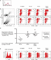
IFN-γ, IL-8 and CD107 as read-outs of freshly isolated decidual leucocyte function. Decidual leucocytes from normal first trimester pregnancies were analysed immediately after isolation by intracellular flow cytometry for the production of IFN-γ (A) or IL-8 (B). Degranulation was assessed after 4 h of culture with HLA class I-negative target cells (C). Decidual leucocytes were gated by scatter and identified by labelling of CD56 or HLA-DR. Compared with an isotype control, little staining for IFN-γ is observed without PMA stimulation (A). The anti-IL-8 mAb stained some freshly isolated decidual leucocytes, but these were HLA-DR+ not CD56+ cells (B). Representative staining for surface CD107a expression by CD56+ NK cells cultured alone and with HLA class I-null 721.221 target cells is shown for dNK cells freshly isolated or first stimulated with IL-15 for 3 days (C). The proportion of CD56+ cells that become CD107a+ after culture with 721.221 targets is then shown for 20 independent donors (D). CD107 up-regulation is quantified by subtracting the CD107 staining detected on NK cells cultured without target cells from that of cultures with target cells.
Figure 5
Figure 5
dNK cell degranulation is not inhibited by target cells expressing different conformations of HLA-G. 721.221 cells transfected with HLA-G (green) or a C42S mutant unable to dimerize (red) were sorted for identical expression levels and compared with untransfected 721.221 cells (grey), monitored by mAb MEM-G/9 staining (A). Lymphocyte preparations were cultured for 3 days with IL-15 and then degranulation of NK cells was detected using flow cytometry for CD107a expression in response to 721.221 target cells expressing HLA-G in monomeric or dimeric conformations. Representative staining is shown of peripheral leucocytes (B) or decidual leucocytes from normal first trimester pregnancies (C). Modulation of NK cell degranulation by the presence of HLA-G is shown for 17 independent donors (D). When the LILRB1+ dNK cells are specifically identified, there is still no evidence for either form of HLA-G-inhibiting degranulation (E).

Similar articles

Cited by

References

    1. Apps R, Gardner L, Sharkey AM, Holmes N, Moffett A. A homodimeric complex of HLA-G on normal trophoblast cells modulates antigen-presenting cells via LILRB1. Eur J Immunol. 2007;37:1924–1937. - PMC - PubMed
    1. Apps R, Gardner L, Moffett A. A critical look at HLA-G. Trends Immunol. 2008;29:313–321. - PubMed
    1. Apps R, Murphy SP, Fernando R, Gardner L, Ahad T, Moffett A. Human leucocyte antigen (HLA) expression of primary trophoblast cells and placental cell lines, determined using single antigen beads to characterize allotype specificities of anti-HLA antibodies. Immunology. 2009;127:26–39. - PMC - PubMed
    1. Ashkar AA, Croy BA. Functions of uterine natural killer cells are mediated by interferon gamma production during murine pregnancy. Semin Immunol. 2001;13:235–241. - PubMed
    1. Banham AH, Colonna M, Cella M, Micklem KJ, Pulford K, Willis AC, Mason DY. Identification of the CD85 antigen as ILT2, an inhibitory MHC class I receptor of the immunoglobulin superfamily. J Leukoc Biol. 1999;65:841–845. - PubMed

Publication types

MeSH terms