. 2011 Jun 10;286(23):20788-96.
doi: 10.1074/jbc.M111.232678. Epub 2011 Apr 6.

Solution structure of the monovalent lectin microvirin in complex with Man(alpha)(1-2)Man provides a basis for anti-HIV activity with low toxicity

Affiliations

Solution structure of the monovalent lectin microvirin in complex with Man(alpha)(1-2)Man provides a basis for anti-HIV activity with low toxicity

Syed Shahzad-ul-Hussan et al. J Biol Chem. .

Abstract

Lectins that bind surface envelope glycoprotein gp120 of HIV with high avidity can potently inhibit viral entry. Yet properties such as multivalency that facilitate strong interactions can also cause nonspecific binding and toxicity. The cyanobacterial lectin microvirin (MVN) is unusual as it potently inhibits HIV-1 with negligible toxicity compared with cyanovirin-N (CVN), its well studied antiviral homolog. To understand the structural and mechanistic basis for these differences, we solved the solution structure of MVN free and in complex with its ligand Manα(1-2)Man, and we compared specificity and time windows of inhibition with CVN and Manα(1-2)Man-specific mAb 2G12. We show by NMR and analytical ultracentrifugation that MVN is monomeric in solution, and we demonstrate by NMR that Manα(1-2)Man-terminating carbohydrates interact with a single carbohydrate-binding site. Synchronized infectivity assays show that 2G12, MVN, and CVN inhibit entry with distinct kinetics. Despite shared specificity for Manα(1-2)Man termini, combinations of the inhibitors are synergistic suggesting they recognize discrete glycans and/or dynamic glycan conformations on gp120. Entry assays employing amphotropic viruses show that MVN is inactive, whereas CVN potently inhibits both. In addition to demonstrating that HIV-1 can be inhibited through monovalent interactions, given the similarity of the carbohydrate-binding site common to MVN and CVN, these data suggest that gp120 behaves as a clustered glycan epitope and that multivalent-protein interactions achievable with CVN but not MVN are required for inhibition of some viruses.


Figures

FIGURE 1.
FIGURE 1.
Analytical ultracentrifugation and NMR relaxation measurements. A, sedimentation velocity data performed with solutions of uniformly labeled 15N-MVN at 60 (red), 30 (blue), and 15 (green) μm. Best fits of the data yield a molecular mass of 12.7 kDa consistent with a monomer. B, longitudinal (T1) and transverse (T2) relaxation data as a function of residue. The rotational correlation time, calculated from the average 15N T1/T2 ratio (excluding the C-terminal mobile residues), is 6.7 ± 0.3 ns.
FIGURE 2.
FIGURE 2.
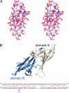
Solution structure of MVN. A, superposition of the final ensemble of 40 simulated annealing structures. None exhibited distance or dihedral angle violations greater than 0.2 Å and 5°, respectively (Table 1). B, ribbon diagram of the restrained minimized mean structure of MVN with domain A colored white (β-strands β4–8) and domain B blue (β-strands β1–3 and β9–10). Disulfide bonds are rendered as gold sticks. Alignment of domains A and B appears below. Coordinates have been deposited to the Protein Data Bank, accession ID 2y1s.
FIGURE 3.
FIGURE 3.
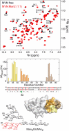
Carbohydrate recognition by MVN. A, superimposed 1H-15N HSQC spectra of free MVN (black) and a 1:1 complex of MVN:Manα(1–2)Man (red). Residues undergoing chemical shift perturbations upon carbohydrate binding are labeled. B, magnitude of the ΔδH/N values as a function of residue showing all resonances to be located in domain A of MVN. ΔδH/N = [(Δ15N2 + Δ1HN2)/2]1/2 in Hz. Sequence alignment by domains appears below, with no perturbations observed for domain B. C, surface representation of MVN with regions colored as in B. D, chemical structure of Man9GlcNAc2. The D1 arm, colored red, includes the model di- and trisaccharides Manα(1–2)Man and Manα(1–2)Manα(1–2)Man.
FIGURE 4.
FIGURE 4.
Solution structure of MVN in complex with Manα(1–2)Man. A, sample planes from a three-dimensional 12C-filtered/13C-separated NOE spectrum showing strong intermolecular NOEs between 13C-attached Asn-55 Hβ and Met-83 Hϵ protons in MVN and 12C-attached protons of α-mannobiose. B, close up of the carbohydrate-binding site with MVN shown as a surface representation and the ligand as sticks. Acidic, polar, and hydrophobic residues comprising the binding site on MVN are colored red, sky blue, and green, respectively. The reducing and terminal mannopyranose rings are labeled A and B, respectively. C, bond rendering showing detailed interactions between MVN and α-mannobiose. Black dotted lines connect atoms located within hydrogen bonding distance; green dashed lines denote van der Waals surfaces. See supplemental Table S2 for structure statistics of complex. Coordinates have been deposited to the Protein Data Bank, accession no. ID 2yhh.
FIGURE 5.
FIGURE 5.
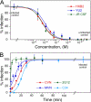
Antiviral activity of MVN in an Env-pseudotyped HIV neutralization assay. A, representative dose-response curves for antiviral activity of MVN against HIV pseudotyped with Env of diverse laboratory-adapted subtype B HIV strains (Table 3). B, infectivity as a function of time of addition of fusion inhibitors in a synchronized HXB2 Env-pseudotyped HIV infection assay. Fully inhibitory concentrations of MVN, CVN, 2G12, and C34 were added at the indicated time points. The experimental data (average of a minimum of four experiments) were fit to a sigmoidal curve (see “Experimental Procedures”). t½ values of the inhibitor-sensitive state are as follows: 2G12 ≤1 min, MVN 8.4 ± 1.6 min, CVN 15.7 ± 1.0 min, C34 21.7 ± 0.6 min.
FIGURE 6.
FIGURE 6.
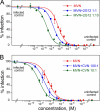
MVN is synergistic with 2G12 (A) and CVN (B). Combination ratios are shown in the figure and listed in Table 3, and combination indices are summarized in Table 4 and supplemental Table S3.

Similar articles

Cited by

References

    1. Taiwo B., Hicks C., Eron J. (2010) J. Antimicrob. Chemother. 65, 1100–1107 - PubMed
    1. Folkers G. K., Fauci A. S. (2010) JAMA 304, 350–351 - PubMed
    1. Balzarini J. (2007) Nat. Rev. Microbiol. 5, 583–597 - PMC - PubMed
    1. Chan D. C., Kim P. S. (1998) Cell 93, 681–684 - PubMed
    1. Leonard C. K., Spellman M. W., Riddle L., Harris R. J., Thomas J. N., Gregory T. J. (1990) J. Biol. Chem. 265, 10373–10382 - PubMed

Publication types

MeSH terms

Associated data