. 2011 May 27;286(21):18775-83.
doi: 10.1074/jbc.M111.230185. Epub 2011 Apr 6.

A disulfide bridge network within the soluble periplasmic domain determines structure and function of the outer membrane protein RCSF

Affiliations

A disulfide bridge network within the soluble periplasmic domain determines structure and function of the outer membrane protein RCSF

Vladimir V Rogov et al. J Biol Chem. .

Abstract

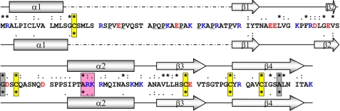
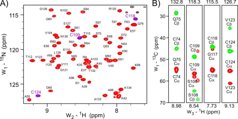
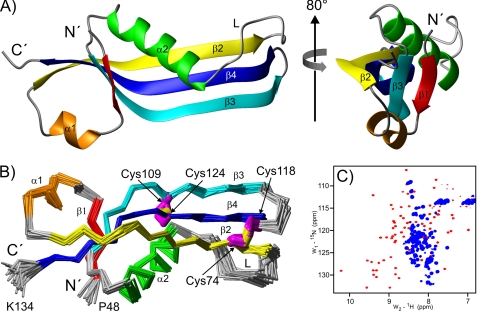
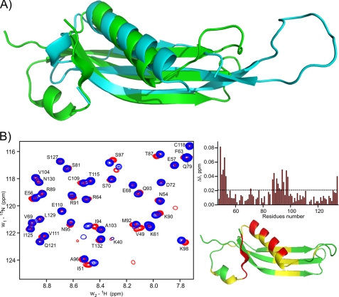
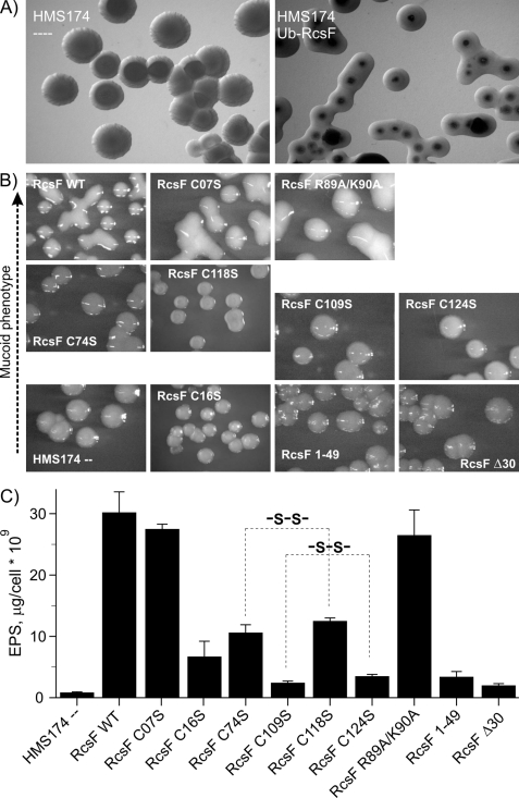
RcsF, a proposed auxiliary regulator of the regulation of capsule synthesis (rcs) phosphorelay system, is a key element for understanding the RcsC-D-A/B signaling cascade, which is responsible for the regulation of more than 100 genes and is involved in cell division, motility, biofilm formation, and virulence. The RcsC-D-A/B system is one of the most complex bacterial signal transduction pathways, consisting of several membrane-bound and soluble proteins. RcsF is a lipoprotein attached to the outer membrane and plays an important role in activating the RcsC-d-A/B pathway. The exact mechanism of activation of the rcs phosphorelay by RcsF, however, remains unknown. We have analyzed the sequence of RcsF and identified three structural elements: 1) an N-terminal membrane-anchored helix (residues 3-13), 2) a loop (residues 14-48), and 3) a C-terminal folded domain (residues 49-134). We have determined the structure of this C-terminal domain and started to investigate its interaction with potential partners. Important features of its structure are two disulfide bridges between Cys-74 and Cys-118 and between Cys-109 and Cys-124. To evaluate the importance of this RcsF disulfide bridge network in vivo, we have examined the ability of the full-length protein and of specific Cys mutants to initiate the rcs signaling cascade. The results indicate that the Cys-74/Cys-118 and the Cys-109/Cys-124 residues correlate pairwise with the activity of RcsF. Interaction studies showed a weak interaction with an RNA hairpin. However, no interaction could be detected with reagents that are believed to activate the rcs phosphorelay, such as lysozyme, glucose, or Zn(2+) ions.


Figures

FIGURE 1.
FIGURE 1.
Sequence alignment and secondary structure prediction for E. coli RcsF. The amino acid sequence of E. coli RcsF (P69411) is shown (positively charged amino acids are shown in blue, negatively charged amino acids in red, and prolines are underlined). The top row of symbols represents conservation within Enterica sequences, and the bottom row represents total conservation between all annotated RcsF sequences. The secondary structure elements predicted by the PsiPred server for E. coli RcsF (P69411) and the most distant annotated RcsF sequence from Aeromonas hydrophilia (A0KMN2) are presented above and below the E. coli sequence, respectively. The boxes emphasize conserved residues (yellow, cysteines; red, positive charged cluster; gray, others).
FIGURE 2.
FIGURE 2.
NMR spectra of RcsF-Δ30 oxidized monomeric form. A, representative area of the RcsF-Δ30 [15N,1H]HSQC-TROSY spectrum. The backbone HN resonances (generally red, HN resonances of cysteines, purple) are almost completely assigned. The non-assigned black contours in the upper right corner belong to Asn/Gln side chains. B, HNCACB strips demonstrating oxidation of all four Δ30 RcsF cysteines. The HN resonance of Cys-74 is not seen in the RcsF-Δ30 TROSY-HSQC spectrum. The positive Cα resonances are shown in red and the negative Cβ resonances in green. Values of >40 ppm for cysteine Cβ resonances indicate an oxidized state.
FIGURE 3.
FIGURE 3.
Structure of the RcsF-Δ30 oxidized monomeric form. A, ribbon diagram representing the RcsF-Δ30 structure. The structure on the right was obtained by 80° rotation around the indicated axis. The unstructured residues 30–47 are not included. B, Cα traces of the final RcsF-Δ30 structural ensemble of 25 conformers. The indicated cysteine side-chains are marked in magenta and the -S–S- bridge in yellow. The structure was obtained from the one shown in A by 180° rotation around the x axis. C, RcsF-Δ30 [15N,1H]HSQC-TROSY spectrum under oxidizing conditions (red contours) and after reduction (blue contours).
FIGURE 4.
FIGURE 4.
Structural similarity of the RcsF-Δ30 oxidized monomeric form to DNA/RNA binding proteins and possible interaction with RNA. A, superimposition of the free L22 protein (cyan, PDB code 1I4J) and RcsF-Δ30 (green). The superimposition with L22 of the Thermus thermophilus 50 S ribosomal subunit (PDB code 2WRJ) is provided in supplemental Fig. 3. B, representative area of the RcsF-Δ30 [15N,1H]HQSC-TROSY spectrum before (red) and after (blue) addition of a 2-fold excess of a 14-mer RNA hairpin (left panel). Right panel, mapping of the observed CSP onto the sequence (top) and the structure (bottom). CSP values below 0.018 ppm (3 × statistical error) are shown in green, those between 0.018–0.036 ppm in yellow, and those with more than 0.036 ppm in red.
FIGURE 5.
FIGURE 5.
Role of the RcsF disulfide bridges in the RcsF-controlled EPS biosynthesis regulation. A, microphotographs of the empty (left panel) and transformed with pET60m_Ub_RcsF WT (right panel) HMS174 cells treated with IPTG and grown at 20 °C. B, microphotographs of HMS174 colonies after IPTG-induced expression of the various RcsF constructs. The pictures are placed in the four rows according to the level of mucoidity of the colonies. The RcsF constructs used in these experiments are indicated in each picture. C, quantification of the polysaccharides production in the HMS174 cells from B. The results of three independent anthrone tests are averaged and expressed in μg of total polysaccharides per 109 cells. Correlated effects of Cys-to-Ser substitutions in the RcsF C-terminal domain are highlighted with dashed lines.

Similar articles

Cited by

References

    1. Majdalani N., Gottesman S. (2005) Annu. Rev. Microbiol. 59, 379–405 - PubMed
    1. Gottesman S., Trisler P., Torres-Cabassa A. (1985) J. Bacteriol. 162, 1111–1119 - PMC - PubMed
    1. Andresen L., Kõiv V., Alamäe T., Mäe A. (2007) FEMS Microbiol. Lett. 273, 229–238 - PubMed
    1. Francez-Charlot A., Laugel B., Van Gemert A., Dubarry N., Wiorowski F., Castanié-Cornet M. P., Gutierrez C., Cam K. (2003) Mol. Microbiol. 49, 823–832 - PubMed
    1. West A. H., Stock A. M. (2001) Trends Biochem. Sci. 26, 369–376 - PubMed

Publication types

MeSH terms

Associated data

LinkOut - more resources