. 2011 Jun 10;286(23):20536-46.
doi: 10.1074/jbc.M110.188326. Epub 2011 Apr 6.

Neisseria meningitidis adhesin NadA targets beta1 integrins: functional similarity to Yersinia invasin

Affiliations

Neisseria meningitidis adhesin NadA targets beta1 integrins: functional similarity to Yersinia invasin

Virginie Nägele et al. J Biol Chem. .

Abstract

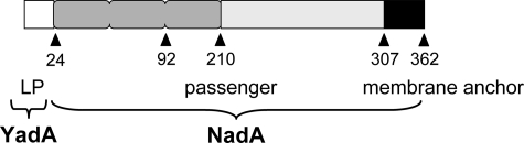
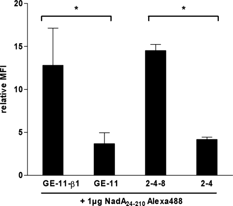
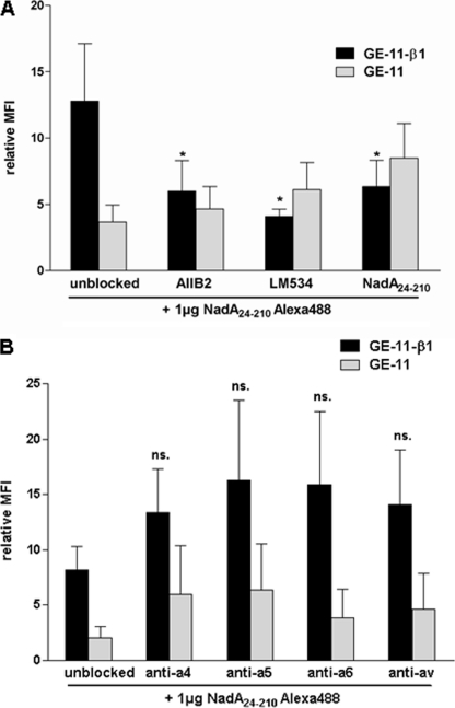
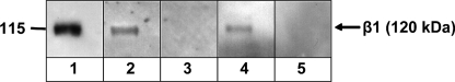
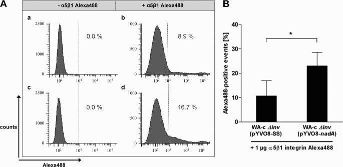
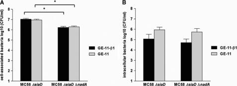
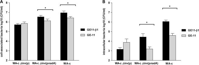
Meningococci are facultative-pathogenic bacteria endowed with a set of adhesins allowing colonization of the human upper respiratory tract, leading to fulminant meningitis and septicemia. The Neisseria adhesin NadA was identified in about 50% of N. meningitidis isolates and is closely related to the Yersinia adhesin YadA, the prototype of the oligomeric coiled-coil adhesin (Oca) family. NadA is known to be involved in cell adhesion, invasion, and induction of proinflammatory cytokines. Because of the enormous diversity of neisserial cell adhesins the analysis of the specific contribution of NadA in meningococcal host interactions is limited. Therefore, we used a non-invasive Y. enterocolitica mutant as carrier to study the role of NadA in host cell interaction. NadA was shown to be efficiently produced and localized in its oligomeric form on the bacterial surface of Y. enterocolitica. Additionally, NadA mediated a β1 integrin-dependent adherence with subsequent internalization of yersiniae by a β1 integrin-positive cell line. Using recombinant NadA(24-210) protein and human and murine β1 integrin-expressing cell lines we could demonstrate the role of the β1 integrin subunit as putative receptor for NadA. Subsequent inhibition assays revealed specific interaction of NadA(24-210) with the human β1 integrin subunit. Cumulatively, these results indicate that Y. enterocolitica is a suitable toolbox system for analysis of the adhesive properties of NadA, revealing strong evidence that β1 integrins are important receptors for NadA. Thus, this study demonstrated for the first time a direct interaction between the Oca-family member NadA and human β1 integrins.


Figures

FIGURE 1.
FIGURE 1.
Schematic representation of full-length NadA protein (allele 1) produced by Y. enterocolitica. The YadA leader peptide (LP), the predicted NadA passenger domain and the membrane anchor domain are shown (alignment according to (4)). Numbers refer to amino acid residues of the protein.
FIGURE 2.
FIGURE 2.
Expression of nadA in Y. enterocolitica. Western blot analysis of outer membrane fractions (10 μg) incubated at 100 °C. A, WA-c Δinv(pnadA) (lane 1); WA-c Δinv(p) (lane 2). B, WA-314 Δyad:nadA (lane 1); WA-314 ΔyadA (lane 2). The assays were performed using rabbit anti-NadA serum and secondary peroxidase-conjugated antibody. The arrows indicate the oligomeric and monomeric form of NadA.
FIGURE 3.
FIGURE 3.
Surface localization of NadA in Y. enterocolitica. Immunofluorescence microscopy showing localization of NadA on the surface of Y. enterocolitica. A, WA-314 ΔyadA:nadA; B, WA-314 ΔyadA (negative control); C, WA-c Δinv(pnadA); D, WA-c Δinv(p) (negative control). Unfixed bacteria were incubated with rabbit anti-NadA serum and secondary anti-rabbit FITC antibody. Scale bar, 10 μm.
FIGURE 4.
FIGURE 4.
Role of nadA-expressing yersiniae in adhesion and invasion into Chang cells. Chang cell monolayers were infected with WA-c Δinv(p) (negative control), WA-c Δinv(pyadA), and WA-c Δinv(pnadA) for 3 h (moi 100). Shown are (A) total cell-associated bacteria (including both intra- and extracellular bacteria), and (B) intracellular bacteria determined using gentamicin protection assays. The number of cell-associated and intracellular bacteria is expressed as log10 colony forming units per ml (CFU/ml). Data are expressed as the means ± S.E. of the mean of at least three independent experiments. *, p < 0.0383 versus WA-c Δinv(p).
FIGURE 5.
FIGURE 5.
Interaction of Alexa488-labeled NadA24–210 protein with epithelial-like and fibroblast-like mouse cells. Epithelial-like GE-11 (human β1 integrin-negative) and GE-11-β1 (human β1 integrin-positive) cells and fibroblast-like 2-4-8 (murine β1 integrin-positive), and 2-4 (murine β1 integrin-negative) cells were incubated with Alexa488-labeled NadA24–210 protein (1 μg/3 × 105 cells) for 1 h at 4 °C and analyzed by flow cytometry. The MFI was related to untreated cells. Data are expressed as the means ± S.E. of at least three independent experiments. *, p < 0.0055.
FIGURE 6.
FIGURE 6.
Inhibition of interaction of Alexa488-labeled NadA24–210 protein with GE-11-β1 cells by using anti-β1 integrin antibodies, unlabeled NadA24–210 protein and anti-α integrin antibodies. Epithelial-like GE-11 (human β1 integrin-negative) and GE-11-β1 (human β1 integrin-positive) cells were treated with indicated β1 integrin specific antibodies (AIIB2 1:4; LM534 1:200) or unlabeled NadA24–210 protein (5 μg/3 × 105 cells) (A), or with α4, α5, α6, or αv integrin specific antibodies (1 μg α4: MAB16983Z, α5: MAB1956Z, αv:MAB1953Z, α6: MAB1378) (B) prior to incubation with Alexa488-labeled NadA24–210 protein (1 μg/3 × 105 cells) and analyzed by flow cytometry. The MFI was related to untreated cells. Data are expressed as the means ± S.E. of at least three independent experiments. *, p < 0.0462, versus unblocked control values.
FIGURE 7.
FIGURE 7.
Far Western blotting of human α5β1 integrin and recombinant NadA24–210 protein. 1 μg of human α5β1 integrin was separated by SDS-PAGE and transferred onto nitrocellulose membranes. Integrins were renatured and detected with anti-β1 integrin antibody (lane 1), NadA specific antibody (lane 3), or Invasin specific antibody (lane 5) and peroxidase-conjugated secondary antibody. Additionally, renatured integrins were incubated with 5 μg of NadA24–210 protein and detected with anti-NadA serum and peroxidase-conjugated secondary antibody (lane 2), or 5 μg of Inv397 (O:8) protein and detected with anti-Invasin serum and peroxidase-conjugated secondary antibody (lane 4). The arrow indicates the size of the β1 integrin subunit at 120 kDa.
FIGURE 8.
FIGURE 8.
Binding of Alexa488-labeled human α5β1 integrin to nadA-expressing yersiniae. 5 × 10⁵ WA-c Δinv(pYV-nadA) (NadA-positive) or WA-c Δinv(pYV-SS) (NadA-negative, negative control) yersiniae were incubated with or without 1 μg of Alexa488-labeled human α5β1 integrin in PBS-0.2% Triton X-100 and 2 mm MnCl2 (5 μg/ml) for 1 h at 4 °C and analyzed by flow cytometry. A, histograms display the result from one representative experiment. a, WA-c Δinv(pYVO8-SS) without addition of Alexa488-labeled α5β1 integrin. b, WA-c Δinv(pYVO8-SS) with addition of Alexa488-labeled α5β1 integrin. c, WA-c Δinv(pYVO8-nadA) without addition of Alexa488-labeled α5β1 integrin. d, WA-c Δinv(pYVO8-nadA) with addition of Alexa488-labeled α5β1 integrin. Percents of Alexa488-positive events are displayed. B, Alexa488-positive events are expressed as the means ± S.E. of four independent experiments. *, p < 0.0416.
FIGURE 9.
FIGURE 9.
Role of NadA in adhesion and invasion of N. meningitidis. For determination of cell-associated bacteria GE-11-β1 and GE-11 cell monolayers were infected for 3 h (moi 100) with unencapsulated N. meningitidis strain MC58 ΔsiaD and the isogenic nadA mutant MC58 ΔsiaD ΔnadA. For determination of intracellular bacteria GE-11-β1 and GE-11 cell monolayers were additionally treated with gentamicin. Shown are (A) total cell-associated bacteria (including both intra- and extracellular bacteria), and (B) intracellular bacteria determined using gentamicin protection assays. The number of cell-associated and intracellular bacteria is expressed as log10 colony forming units per ml (CFU/ml). Data are expressed as the means ± S.E. of the mean of at least three independent experiments. *, p < 0.0001.
FIGURE 10.
FIGURE 10.
Role of nadA-expressing yersiniae in adhesion and invasion. GE-11-β1 and GE-11 cell monolayers were infected with WA-c Δinv(p) (negative control), WA-c Δinv(pnadA), and WA-c (positive control) for 1 h (moi 50). Shown are (A) total cell-associated bacteria (including both intra- and extracellular bacteria), and (B) intracellular bacteria determined using gentamicin protection assays. The number of cell-associated and intracellular bacteria is expressed as log10 colony forming units per ml (CFU/ml). Data are expressed as the means ± S.E. of the mean of at least three independent experiments. *, p < 0.041.

References

    1. Virji M. (2009) Nat. Rev. Microbiol. 7, 274–286 - PubMed
    1. Cotter S. E., Surana N. K., St. Geme J. W., 3rd (2005) Trends Microbiol. 13, 199–205 - PubMed
    1. Henderson I. R., Navarro-Garcia F., Desvaux M., Fernandez R. C., Ala'Aldeen D. (2004) Microbiol. Mol. Biol. Rev. 68, 692–744 - PMC - PubMed
    1. Hoiczyk E., Roggenkamp A., Reichenbecher M., Lupas A., Heesemann J. (2000) EMBO J. 19, 5989–5999 - PMC - PubMed
    1. Linke D., Riess T., Autenrieth I. B., Lupas A., Kempf V. A. (2006) Trends Microbiol. 14, 264–270 - PubMed

Publication types

LinkOut - more resources