. 2011 Jun 3;286(22):19179-90.
doi: 10.1074/jbc.M110.200790. Epub 2011 Apr 6.

Differential use of chondroitin sulfate to regulate hyaluronan binding by receptor CD44 in Inflammatory and Interleukin 4-activated Macrophages

Affiliations

Differential use of chondroitin sulfate to regulate hyaluronan binding by receptor CD44 in Inflammatory and Interleukin 4-activated Macrophages

Brian Ruffell et al. J Biol Chem. .

Abstract

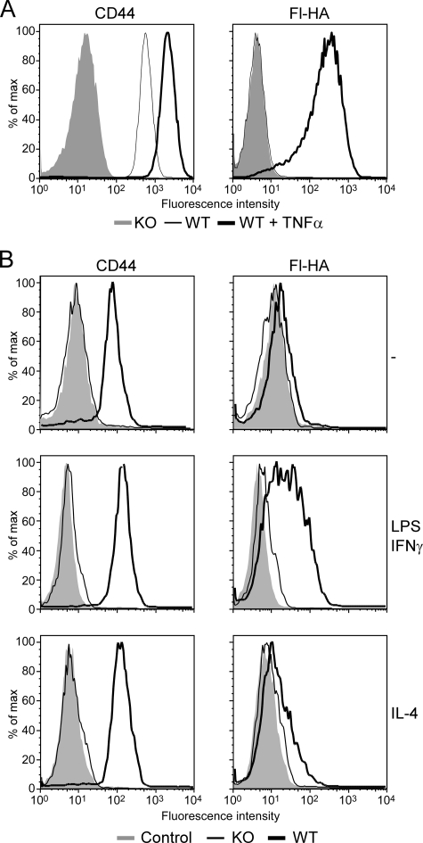
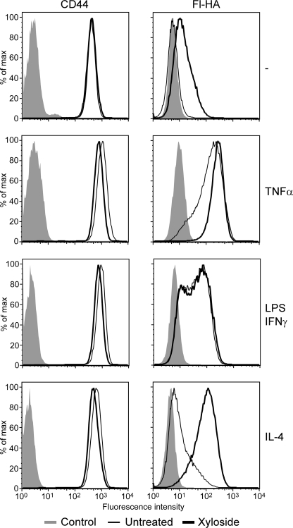
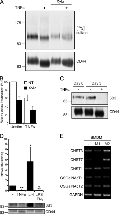
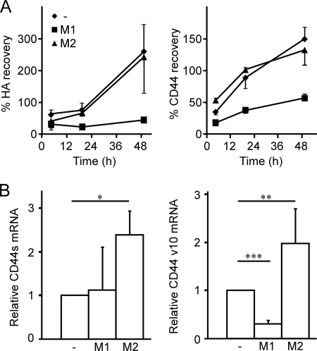
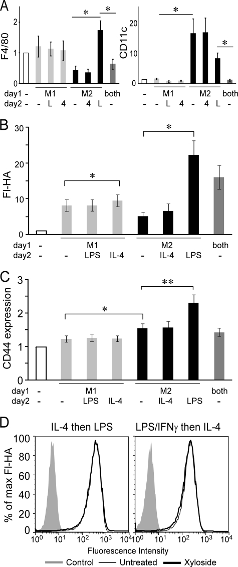
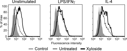
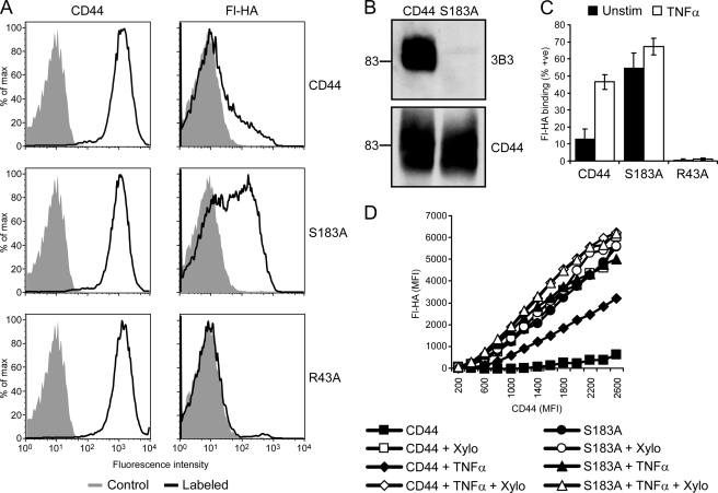
CD44 is a cell surface receptor for the extracellular matrix glycosaminoglycan hyaluronan and is involved in processes ranging from leukocyte recruitment to wound healing. In the immune system, the binding of hyaluronan to CD44 is tightly regulated, and exposure of human peripheral blood monocytes to inflammatory stimuli increases CD44 expression and induces hyaluronan binding. Here we sought to understand how mouse macrophages regulate hyaluronan binding upon inflammatory and anti-inflammatory stimuli. Mouse bone marrow-derived macrophages stimulated with tumor necrosis factor α or lipopolysaccharide and interferon-γ (LPS/IFNγ) induced hyaluronan binding by up-regulating CD44 and down-regulating chondroitin sulfation on CD44. Hyaluronan binding was induced to a lesser extent in interleukin-4 (IL-4)-activated macrophages despite increased CD44 expression, and this was attributable to increased chondroitin sulfation on CD44, as treatment with β-d-xyloside to prevent chondroitin sulfate addition significantly enhanced hyaluronan binding. These changes in the chondroitin sulfation of CD44 were associated with changes in mRNA expression of two chondroitin sulfotransferases, CHST3 and CHST7, which were decreased in LPS/IFNγ-stimulated macrophages and increased in IL-4-stimulated macrophages. Thus, inflammatory and anti-inflammatory stimuli differentially regulate the chondroitin sulfation of CD44, which is a dynamic physiological regulator of hyaluronan binding by CD44 in mouse macrophages.


Figures

FIGURE 1.
FIGURE 1.
Induction of hyaluronan binding and CD44 in bone marrow-derived macrophages. CD44 expression and hyaluronan binding were analyzed by flow cytometry in bone marrow-derived macrophages derived from wild type or CD44−/− mice and stimulated with 20 ng/ml TNFα (A) or 100 ng/ml LPS and 50 ng/ml IFN-γ or with 10 ng/ml IL-4 (B) for 2 days. A, the left panel shows expression levels of CD44, detected using Alexa 647 conjugated IM7, from unstimulated (dashed line) and TNFα-stimulated (thick line) bone marrow-derived macrophages, whereas the right panel shows binding to fluorescent-hyaluronan (Fl-HA). CD44 and Fl-HA binding of CD44−/− bone marrow-derived macrophages is shown by the shaded histograms. B, CD44 expression levels were detected using Pacific blue-conjugated IM7 on unstimulated, LPS/IFNγ stimulated, and IL-4 stimulated bone marrow-derived macrophages derived from wild type (thick line) and CD44−/− mice (thin line). Negative controls (shaded histograms) are unlabeled cells. This is one representative experiment repeated at least three times.
FIGURE 2.
FIGURE 2.
TNFα does not increase CD44 sulfation in bone marrow-derived macrophages, and chondroitin sulfate-modified CD44 negatively correlates with hyaluronan binding in human KG1a cells. A, shown is an autoradiograph of CD44 immunoprecipitated from sodium [35S]sulfate-labeled bone marrow-derived macrophages and resolved by SDS-PAGE. CD44 loading (lower panel) was determined by Western blotting with J1WBB. B, CHST2 (GlcNAc6ST-1) and CHST7 (GlcNAc6ST-4) mRNA expression from unstimulated and TNFα-stimulated bone marrow-derived macrophages (BMDM) is shown. Isolated mRNA was subjected to semiquantitative PCR, and β-actin expression was used as a loading control. RNA isolated from mouse brain or kidney was used as a positive control for CHST2 or CHST7, respectively. C, shown is flow cytometry of CD44 expression and fluorescent-hyaluronan (Fl-HA) binding by KG1a cells sorted for either low or high hyaluronan binding. D, detection of chondroitin sulfate on immunoprecipitated CD44 by Western blotting with the anti- 4-sulfated chondroitin sulfate stub mAb, 2B6 is shown. CD44 loading was determined by blotting with the anti-human mAb 3G12 (lower panel). E, densitometry was performed on the Western blots from D, and after controlling for differences in CD44 loading, the amount of 2B6 detected was set to 1 for the high hyaluronan binding cells. Data are the mean ± S.D. of four experiments, and statistical significance (**p < 0.01) is shown compared with low cells.
FIGURE 3.
FIGURE 3.
Inhibition of glycosaminoglycan addition by β-d-xyloside increases hyaluronan binding by bone marrow-derived macrophages. CD44 expression and hyaluronan binding were analyzed by flow cytometry in unstimulated bone marrow-derived macrophages or bone marrow-derived macrophages stimulated with 20 ng/ml TNFα, 100 ng/ml LPS and 10 ng/ml IFNγ, or 10 ng/ml IL-4 for 2 days in the presence (thick line) or absence (thin line) of 2 mm β-d-xyloside. The left panel shows expression levels of CD44, detected using Alexa 647-conjugated IM7, whereas the right panel shows binding to fluorescent-hyaluronan (Fl-HA). Unlabeled cells (shaded histograms) were used as negative controls. This is one representative experiment repeated three times.
FIGURE 4.
FIGURE 4.
Chondroitin sulfate addition to CD44 is reduced by inflammatory cytokines. A, shown is an autoradiograph of CD44 immunoprecipitated from sodium [35S]sulfate-labeled bone marrow-derived macrophages grown in the presence or absence of 2 mm β-d-xyloside (Xylo) and resolved by SDS-PAGE. CD44 loading was determined by Western blotting with J1WBB. B, relative sulfate incorporation by CD44 as measured by densitometry is shown. Sulfate incorporation per unit CD44 was assessed and set to 100% in unstimulated, non-treated (NT) cells. Values represent the mean ± S.D. of six independent experiments. C, detection of chondroitin sulfate on immunoprecipitated CD44 from unstimulated and TNFα-stimulated bone marrow-derived macrophages by Western blotting with the anti-6-sulfated chondroitin sulfate stub mAb, 3B3, is shown. CD44 loading levels were determined by Western blotting with the CD44 antiserum, J1WBB. D, analysis of chondroitin sulfate addition to CD44 from bone marrow-derived macrophages stimulated with TNFα, IL-4, or with LPS and IFNγ. Western blots (a representative is shown in the bottom panel) were analyzed by densitometry, and after taking into account variations in CD44 loading, the intensity of 3B3 staining for CD44 from unstimulated bone marrow-derived macrophages was set to 1. Data are shown as the mean ± S.D. of 3 experiments with significance indicated as: *, p < 0.05; **, p < 0.01; ***, p < 0.001. E, CHST3 (C6ST1), CHST7 (C6ST2), CHST1 (KSGal6ST), CSGalNAcT1, and CSGalNAcT2 mRNA expression from unstimulated (−), 100 ng/ml LPS and 50 ng/ml IFNγ-stimulated (M1), and 10 ng/ml IL-4-stimulated (M2) bone marrow-derived macrophages is shown. Isolated mRNA was subjected to semiquantitative PCR, and GAPDH expression was used as a loading control. These are representative data that was reproduced with macrophages from four mice.
FIGURE 5.
FIGURE 5.
Macrophage polarization affects CD44 turnover and expression of variable exon 10. A, percent recovery of fluorescent-hyaluronan (HA) binding and CD44 expression after trypsin treatment of unstimulated, M1, and M2 bone marrow-derived macrophages is shown. Bone marrow-derived macrophages were unstimulated (−) or stimulated with either 100 ng/ml LPS and 50 ng/ml IFNγ (M1) or 10 ng/ml IL-4 (M2) for 1 day. Cells were then treated with trypsin for 5 min or untreated, then further cultured for 3, 18, and 48 h. The percent recovery was calculated from flow cytometry, by dividing the mean fluorescence value of fluorescent-hyaluronan or CD44 from the trypsin-treated samples by the value of the non-trypsin treated cells at each time point. Data shown are the mean ± S.D. of three biological replicates. B, relative mRNA expression of CD44s and CD44v10 in bone marrow-derived macrophages is shown. Bone marrow-derived macrophages were cultured in media (−) or stimulated for 48 h with 50 ng/ml IFNγ and 100 ng/ml LPS (M1) or 10 ng/ml IL-4 (M2). Total mRNA was extracted and subjected to reverse transcription. CD44s (left panel) and CD44v10 (right panel) transcripts were measured by quantitative-PCR and normalized to GAPDH expression. Expression is shown relative to the unstimulated control. Graphs show the average relative expression from five mice over three experiments ± S.D. with significance indicated as p < 0.05 (*), p < 0.01 (**), and p < 0.001 (***).
FIGURE 6.
FIGURE 6.
Distinct phenotypes of classically activated (M1) and alternatively activated (M2) bone marrow-derived macrophages and the relative expression levels of CD44 and fluorescent-hyaluronan binding. A–C, bone marrow-derived macrophages were stimulated with either 100 ng/ml LPS and 50 ng/ml IFN-γ (M1), 10 ng/ml IL-4 (M2), 100 ng/ml LPS, 50 ng/ml IFN-γ, and 10 ng/ml IL-4 (both) or media control (−) on day 1, then re-stimulated with LPS (L), IL-4 (4) or media alone (−) on day 2. Graphs show the mean fluorescent intensity ± S.E. of at least three experiments with significance indicated as: *, p < 0.05; **, p < 0.01. A, shown is the relative increase in expression levels of F4/80 (left panel) and CD11c (right panel) of activated versus unstimulated bone marrow-derived macrophages. B and C, shown is the relative increase in fluorescent-hyaluronan (Fl-HA) binding (B) and CD44 expression levels (C) compared with unstimulated bone marrow-derived macrophages. D, bone marrow-derived macrophages were stimulated in the presence (black line) or absence (thin line) of 2 mm β-d-xyloside, and the negative control (shaded histogram) was unlabeled cells. Cells were stimulated with IL-4 followed by LPS on the second day (IL-4 then LPS) or stimulated with LPS and IFN-γ followed by IL-4 on the second day (LPS/IFN-γ then IL-4). One of four experiments is shown.
FIGURE 7.
FIGURE 7.
Effect of inhibition of glycosaminoglycan addition on hyaluronan binding by peritoneal macrophages. Fluorescent-hyaluronan (Fl-HA) binding was analyzed by flow cytometry after gating on F4/80-positive macrophages. These peritoneal macrophages were stimulated ex vivo with 20 ng/ml IL-4 or 100 ng/ml LPS and 10 ng/ml IFNγ for 2 days in the presence (thick line) or absence (thin line) of 2 mm β-d-xyloside. Unlabeled cells (shaded histograms) were used as negative controls. One of four experiments is shown.
FIGURE 8.
FIGURE 8.
Chondroitin sulfate addition to CD44 regulates hyaluronan binding in bone marrow-derived macrophages. During the generation of bone marrow-derived macrophages, bone marrow from CD44−/− mice was infected with a retrovirus expressing the YFP marker and wild type CD44, S183A mutant CD44, or R43A mutant CD44. A, shown is CD44 expression and fluorescent-hyaluronan (Fl-HA) binding by YFP-positive cells, with unlabeled cells as negative controls (shaded histogram). B, shown is detection of chondroitin sulfate on immunoprecipitated CD44 from CD44 and S183A-CD44 infected bone marrow-derived macrophages by Western blotting with the anti-chondroitin sulfate mAb 3B3. CD44 levels were determined using J1WBB antiserum. C, shown is the percent of fluorescent-hyaluronan binding bone marrow-derived macrophages expressing wild type, S183A mutant CD44, or R43A mutant CD44 after culture in the presence or absence of TNFα for 3 days. Cells were analyzed by flow cytometry after labeling with Alexa 647-conjugated IM7 and fluorescent-hyaluronan. Data are shown as the mean ± S.D. of 5 experiments. D, shown is fluorescent-hyaluronan binding versus CD44 expression in bone marrow-derived macrophages cultured with or without TNFα in the presence or absence of β-d-xyloside (Xylo) for 3 days. Cells were analyzed by flow cytometry as above, and numerous gates were drawn based on CD44 expression levels to determine the mean fluorescent intensity (MFI) for fluorescent-hyaluronan binding in each of these gates. To control for leakage of the YFP signal, the MFI of cells infected with CD44 containing the R43A mutation was subtracted from the original values. The final MFI value for fluorescent-hyaluronan was then plotted against the MFI used for each CD44 gate.

Similar articles

Cited by

References

    1. Zhang X., Mosser D. M. (2008) J. Pathol. 214, 161–178 - PMC - PubMed
    1. Geissmann F., Manz M. G., Jung S., Sieweke M. H., Merad M., Ley K. (2010) Science 327, 656–661 - PMC - PubMed
    1. Marriott H. M., Dockrell D. H. (2007) Exp. Lung Res. 33, 493–505 - PubMed
    1. Van Ginderachter J. A., Movahedi K., Hassanzadeh Ghassabeh G., Meerschaut S., Beschin A., Raes G., De Baetselier P. (2006) Immunobiology 211, 487–501 - PubMed
    1. Gordon S. (2007) Eur. J. Immunol. 37, S9–S17 - PubMed

Publication types

MeSH terms

LinkOut - more resources