. 2011 Jun;85(12):5995-6007.
doi: 10.1128/JVI.00034-11. Epub 2011 Apr 6.

An anti-inflammatory role of VEGFR2/Src kinase inhibitor in herpes simplex virus 1-induced immunopathology

Affiliations

An anti-inflammatory role of VEGFR2/Src kinase inhibitor in herpes simplex virus 1-induced immunopathology

Shalini Sharma et al. J Virol. 2011 Jun.

Abstract

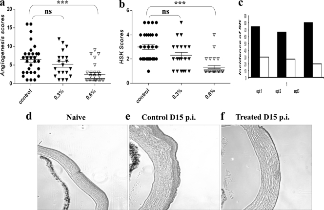
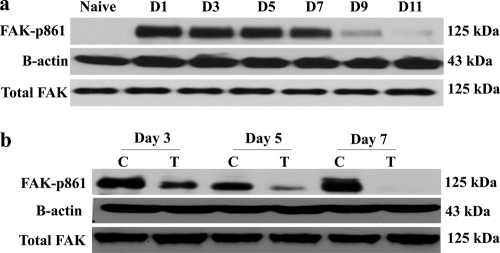
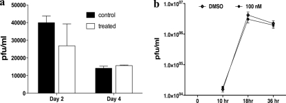
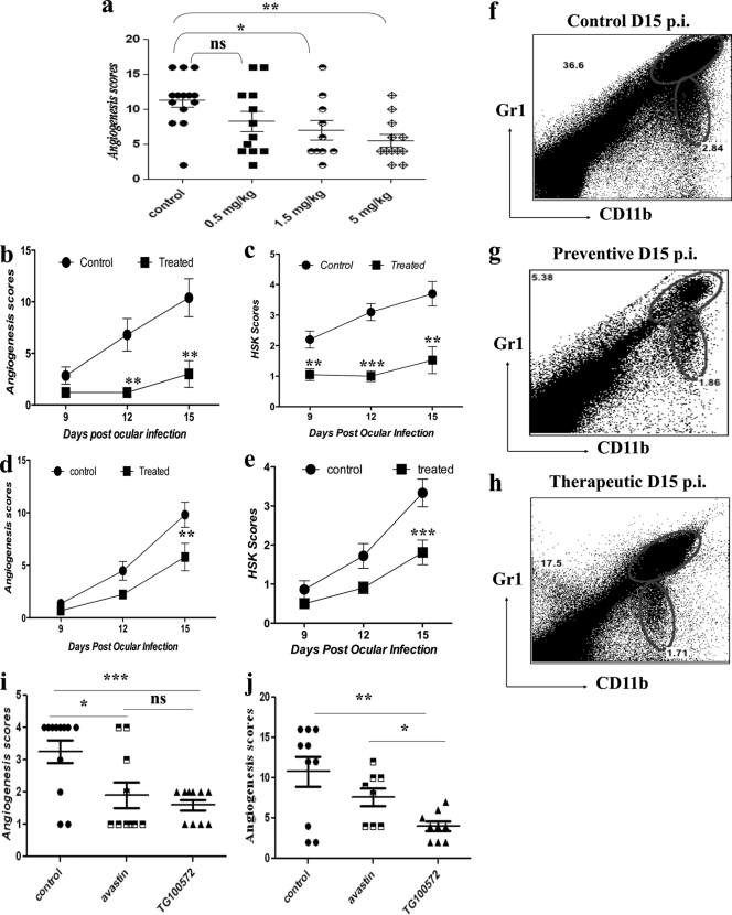
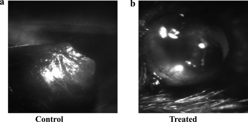
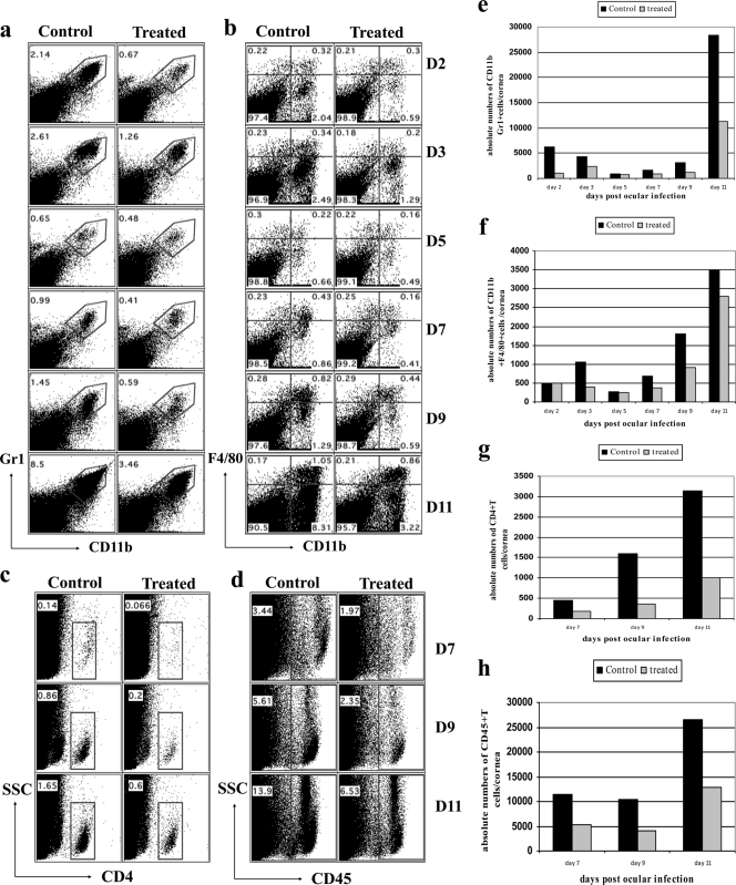
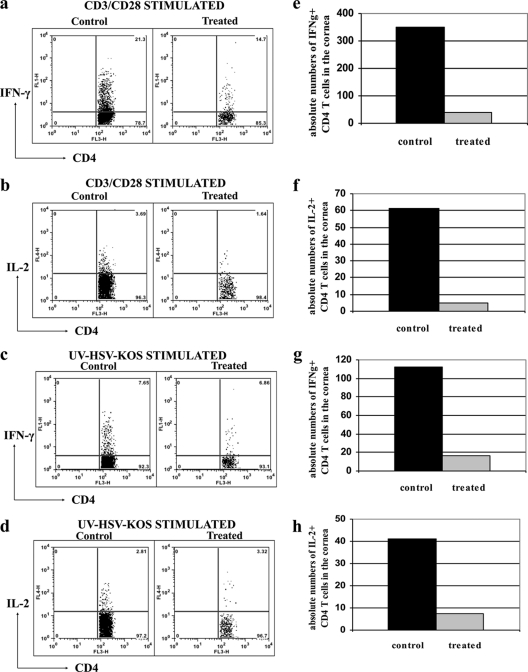
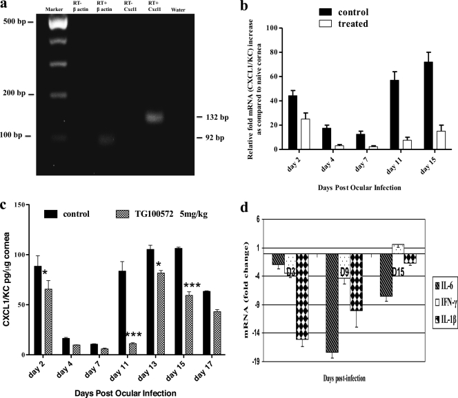
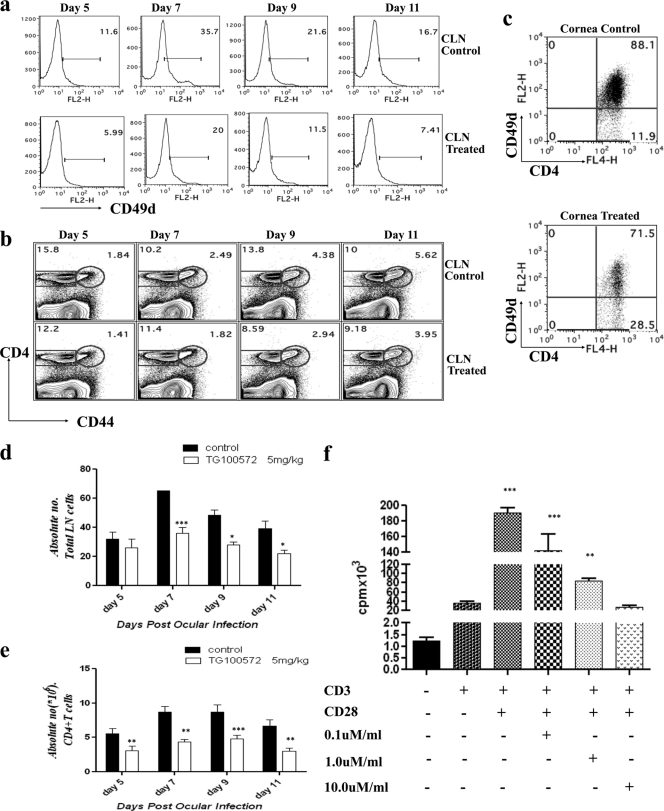
Corneal neovascularization represents a key step in the blinding inflammatory stromal keratitis (SK) lesion caused by ocular infection with herpes simplex virus (HSV). In this report, we describe a novel approach for limiting the angiogenesis caused by HSV infection of the mouse eye. We show that topical or systemic administration of the Src kinase inhibitor (TG100572) that inhibits downstream molecules involved in the vascular endothelial growth factor (VEGF) signaling pathway resulted in markedly diminished levels of HSV-induced angiogenesis and significantly reduced the severity of SK lesions. Multiple mechanisms were involved in the inhibitory effects. These included blockade of IL-8/CXCL1 involved in inflammatory cells recruitment that are a source of VEGF, diminished cellular infiltration in the cornea, and reduced proliferation and migration of CD4(+) T cells into the corneas. As multiple angiogenic factors (VEGF and basic fibroblast growth factor [bFGF]) play a role in promoting angiogenesis during SK and since Src kinases are involved in signaling by many of them, the use of Src kinase inhibition represents a promising way of limiting the severity of SK lesions the most common cause of infectious blindness in the Western world.


Figures

Fig. 1.
Fig. 1.
Effect of topical administration of the Src kinase inhibitor (TG100801) on the severity of SK. C57BL/6 animals infected with 104 PFU of HSV-1 were treated with TG100801 (0.3% and 0.6%, topically). TG100801 was applied topically starting at day 1 and continuing until day 14 postinfection (p.i.). (a, b) Cumulative data for angiogenesis (a) and HSK (b) scores at day 12 p.i. as measured by slit lamp biomicroscopy in three independent experiments. The levels of significance were determined by one-way ANOVA using Dunnett's post hoc setting. ***, P ≤ 0.001. (c) The bar diagram demonstrates the percent severity for each group of mice infected with 104 PFU of HSV-1 RE at day 15 p.i. in three independent experiments. The SK scores of 3 and >3 were counted as representing SK incidence (black bars represent the infected control; white bars represent TG100801-treated animals). (d to f) H&E staining of the corneal sections of naïve (d), control (e), and TG100801-treated (f) animals.
Fig. 2.
Fig. 2.
TG100801 inhibits FAK-y861 phosphorylation in the murine cornea. (a) Western blot analysis for phosphorylated FAK861 in corneal cell lysate at different time points following HSV-1 infection. (b) Inhibition of FAK phosphorylation. Mice were infected with 104 PFU of HSV-1 RE. Twenty four hours following infection, one group was treated topically (cornea) with a 5-μl drop of 0.6% TG100801, twice daily, until day 14 p.i. The second group received the vehicle control in the same way. Corneal cell lysate was used to detect the level of phosphorylated FAKY861 by anti-rabbit FAK antibody in the Western blot. Drug-treated mice showed inhibition of FAK phosphorylation at all time points tested within the periods of elevated phosphorylated FAK. N, naïve; C, control; T, treated.
Fig. 3.
Fig. 3.
TG100572 controls SK lesion severity independent of viral replication. (a) Viral titers in corneal swabs in control and TG100801-treated mice at days 2 and 4 p.i. are shown. (b) Vero cells were infected at an MOI of 1 and incubated with 100 nM TG100572 (maximum below-cytotoxicity level) after infection or with 0.1% DMSO. The effect of the drug (TG100572) on production of HSV-1 was determined by titrating the virus in cell culture supernatant at different time points following infection.
Fig. 4.
Fig. 4.
Effect of systemic administration of Src kinase inhibitor (TG100572) on angiogenesis and SK lesion severity. (a) C57BL/6 animals infected with 104 PFU of HSV were treated with different concentrations of TG100572 (0.5, 1.5, and 5 mg/kg of body weight) intraperitoneally, daily, starting from either 24 h postinfection (preventive) or day 6 p.i. (therapeutic manipulation) and continuing until day 14. The comparative angiogenesis scores obtained at day 14 for HSV-infected animals treated with the indicated concentrations of TG100572 are shown. The statistical significance was determined using one-way ANOVA. (b to e) Kinetics of angiogenesis and lesion expression in control and TG100572-treated animals at 9 to 15 days postinfection (dpi) is shown. Disease progression in control and TG100572-treated mice following infection with 104 PFU of HSV-1 in C57BL/6 animals under preventive (b and c) and therapeutic (d and e) modes of treatment. The level of significance was calculated by two-way ANOVA. (i and j) Comparative angiogenesis scores for bevacizumab and TG100572-treated mice compared to the levels for infected but untreated controls at day 9 p.i. (i) and day 14 p.i. (j). The statistical significance was determined by Student's t test. (f to h) Infiltration of CD11b+ Gr1+ polymorph nuclear neutrophils in the corneas of the control group (f) and following preventive (g) and therapeutic (h) treatment at day 15 p.i. All experiments were repeated at least three times. *, P ≤ 0.05; **, P ≤ 0.01; ***, P ≤ 0.001.
Fig. 5.
Fig. 5.
Representative eye photograph (at day 15 p.i.) of control (a) and TG100572-treated (b) mice.
Fig. 6.
Fig. 6.
Kinetics of cellular infiltration in the corneas of control and TG100572-treated mice. Effects of TG100572 treatment on kinetics of cellular infiltration in corneas of HSV-infected animals analyzed by flow cytometry. C57BL/6 mice infected with 104 PFU of HSV were either treated with TG100572 (5 mg/kg of body weight) i.p. daily starting from 24 h p.i. and continuing until day 14 p.i. or untreated controls. A single-cell suspension of the infected corneas was prepared from 6 pooled corneas (n = 3) at the indicated time points from each group of mice (TG100572 treated or control). The cells were labeled for Gr1+ CD11b+ (polymorph nuclear cells) (a, e), CD11b+ F4/80+ (macrophage) (b, f), CD4+ (c, g), and CD45+ (leukocyte common antigen; pan-leukocyte marker) (d, h). The numbers on the dot plots indicate the percentages of the cells expressing the particular markers in control and kinase inhibitor-treated mice at the indicated time points p.i. The experiment was repeated three times, and data are representative of a single experiment.
Fig. 7.
Fig. 7.
TG100572 treatment diminishes the infiltration of pathogenic Th1 cells in the cornea. C57BL/6 mice infected with 104 PFU of HSV-1 were treated with TG100572 (5 mg/kg of body weight) i.p. daily, staring from 24 h p.i. and continuing until the termination of the experiments. A single-cell suspension of the infected corneas was prepared from 6 pooled corneas at day 15 p.i. from each group of mice (control and treated). The cells were stimulated with either anti-CD3 anti-CD28 or UV-inactivated HSV Kos and stained for CD4+ T cells producing IFN-γ and IL-2. Frequencies (a and b) and absolute numbers (per cornea) (e and f) of IFN-γ+ and IL-2+ T cells, respectively, in the control and treated groups were observed following stimulation by anti-CD3 anti-CD28. The frequencies (c and d) and absolute numbers (g and h) of IFN-γ+ and IL-2+ T cells, respectively, in the control and treated groups following stimulation by UV-inactivated HSV Kos are shown. The experiment was repeated three times, and data are representative of a single experiment.
Fig. 8.
Fig. 8.
TG100572 treatment results in the blockade of CXCL1 in the cornea. (a) Agarose gel analysis for CXCL1 (132 bp) (4th lane) transcripts from infected corneas is shown. The initial lane is a marker; the 2nd lane represents β-actin (92 bp). The 1st and 3rd lanes represent the RT-negative control for β-actin and CXCL1, respectively. The 5th lane represents a negative control (water). (b) Kinetic analysis for the expression of CXCL1 mRNA by qPCR at different time points p.i. after Src kinase inhibitor or mock treatment is shown. Wild-type (WT) mice were infected with HSV and mock treated or treated with TG100572. Six corneas were harvested from the respective groups at the indicated time points, pooled, and subjected to quantification by qPCR for CXCL1 mRNA. (c) Quantification of CXCL1 protein in HSV-1-infected corneas by ELISA after mock or TG100572 treatment is shown. At each time point, 6 corneas were harvested from HSV-infected mice mock treated or treated with TG100572, and levels of CXCL1 protein were determined by ELISA. The level of significance was determined using two-way ANOVA with the Bonferroni posttest. *, P ≤ 0.05; ***, P ≤ 0.001. (d) Reduction in IL-6, IFN-γ, and IL-1β after TG100572 treatment is shown. The mice infected with HSV were mock treated or treated with TG100572, and corneas collected from the respective groups at the indicated time points were subjected to qPCR for IL-6, IFN-γ, and IL-1β mRNAs. The values are represented as fold changes mRNA compared to the level for the infected control. The above-described experiments were repeated three times.
Fig. 9.
Fig. 9.
Src kinase inhibition may result in the attenuation of T cell function. (a) Kinetic analysis of CD49d expression on the CD4+ T cells in the control and treated animals after ocular HSV-1 infection. C57BL/6 mice were infected with 104 PFU of HSV. Mice (n = 3) were sacrificed at each indicated time point, and their draining cervical lymph nodes and corneas were analyzed for surface expression of CD4 and CD49d by flow cytometry. (a) Histograms representing the percentages of CD49d expression on CD4+ T cells in draining cervical lymph nodes at the indicated time points postinfection. Data shown are from one representative experiment. (b) Reduced expression of CD44 on CD4+ T cells in draining CLN of TG100572-treated mice. (c) FACS plots showing the percentages of CD4+ CD49d+ T cells in the corneas in control and TG100572-treated animals at day 11 postinfection. All kinetic experiments were repeated at least twice. (d, e) Absolute numbers (106) of the total lymph node cells (d) and CD4+ T cells (e) in control and treated animals are shown for the indicated time points p.i. The level of significance was determined using two-way ANOVA with the Bonferroni posttest. (f) DLN cells were enriched for CD4+ T cells and stimulated with anti-CD3 anti-CD28 in the presence or absence of drug at the indicated concentrations. Cell proliferation results are expressed as mean cpm from triplicate cultures. *, P ≤ 0.05; **, P ≤ 0.01; ***, P ≤ 0.001.

Similar articles

Cited by

References

    1. Abu-Ghazaleh R., Kabir J., Jia H., Lobo M., Zachary I. 2001. Src mediates stimulation by vascular endothelial growth factor of the phosphorylation of focal adhesion kinase at tyrosine 861, and migration and anti-apoptosis in endothelial cells. Biochem. J. 360:255–264 - PMC - PubMed
    1. Ambati B. K., et al. 2006. Corneal avascularity is due to soluble VEGF receptor-1. Nature 443:993–997 - PMC - PubMed
    1. Biswas P. S., Rouse B. T. 2005. Early events in HSV keratitis—setting the stage for a blinding disease. Microbes Infect. 7:799–810 - PubMed
    1. Bock F., et al. 2007. Bevacizumab as a potent inhibitor of inflammatory corneal angiogenesis and lymphangiogenesis. Invest. Ophthalmol. Vis. Sci. 48:2545–2552 - PubMed
    1. Doukas J., et al. 2008. Topical administration of a multi-targeted kinase inhibitor suppresses choroidal neovascularization and retinal edema. J. Cell. Physiol. 216:29–37 - PMC - PubMed

Publication types

MeSH terms

Substances