. 2011 Jun;85(12):5773-81.
doi: 10.1128/JVI.00039-11. Epub 2011 Apr 6.

Rescue of infectious particles from preassembled alphavirus nucleocapsid cores

Affiliations

Rescue of infectious particles from preassembled alphavirus nucleocapsid cores

Jonathan E Snyder et al. J Virol. 2011 Jun.

Abstract

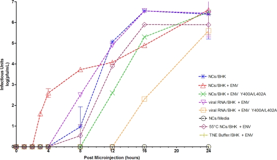
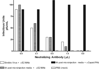
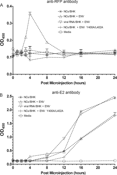
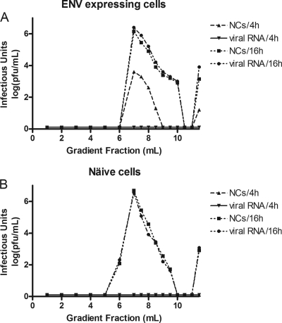
Alphaviruses are small, spherical, enveloped, positive-sense, single-stranded, RNA viruses responsible for considerable human and animal disease. Using microinjection of preassembled cores as a tool, a system has been established to study the assembly and budding process of Sindbis virus, the type member of the alphaviruses. We demonstrate the release of infectious virus-like particles from cells expressing Sindbis virus envelope glycoproteins following microinjection of Sindbis virus nucleocapsids purified from the cytoplasm of infected cells. Furthermore, it is shown that nucleocapsids assembled in vitro mimic those isolated in the cytoplasm of infected cells with respect to their ability to be incorporated into enveloped virions following microinjection. This system allows for the study of the alphavirus budding process independent of an authentic infection and provides a platform to study viral and host requirements for budding.


Figures

Fig. 1.
Fig. 1.
Detection of infectious unit release after microinjection of cells with nucleocapsids. Nucleocapsids (NCs) or viral RNAs were microinjected into the cell type as indicated. Cells expressed wild-type envelope proteins containing an RFP:E2 fusion (BHK + ENV), the envelope proteins containing the E2 Y400A/L402A mutations in addition to the RFP:E2 fusion (BHK + ENV Y400A/L402A), or no viral envelope proteins (BHK). In some cases, NCs were heat shocked at 55°C for 3 min immediately before microinjection. At the time points indicated on the x axis, media were removed from cells and replaced with fresh media. Media were assayed by plaque assay for the presence of infectious units, and the titer [log(PFU/ml)] was plotted as a function of time postmicroinjection. Microinjection of NC buffer (TNE) into BHK cells expressing the envelope proteins represented a mock sample, and NCs microinjected directly into the media provided an additional negative control.
Fig. 2.
Fig. 2.
Neutralization of infectious units released after microinjection of nucleocapsids. The ability of infectious units in the postmicroinjection media to initiate an infection can be reduced by incubation with a known SINV-neutralizing antibody. One hundred PFU of infectious particles (from postmicroinjection media) or purified virus stock was incubated with different volumes of neutralizing antibody anti-E2 MAb, and the resultant infectivity was analyzed by plaque assay. Incubation with nonneutralizing anticapsid antibody, anticapsid PAb, was used as a negative control, and the addition of PBS to cells, rather than infectious media, represents a mock sample.
Fig. 3.
Fig. 3.
ELISA detects release of RFP-containing VLPs after microinjection. Media isolated at various time points after microinjection of cytoplasm-derived NCs into cells expressing the viral envelope proteins were concentrated and applied to 96-well ELISA plates. Plates were probed with either an anti-RFP (DS Red) primary antibody (A) or an anti-E2 primary antibody (B) followed by an HRP-conjugated secondary antibody. The signal was measured optically at 450 nm. Medium isolated from cells expressing the envelope proteins, but not microinjected with NCs, was used as a negative control.
Fig. 4.
Fig. 4.
Gradient analysis of infectious units. Postmicroinjection media (1 ml) were applied to a continuous 10-ml 0 to 30% iodixanol gradient and centrifuged at 32,000 rpm in a Beckman SW-41 rotor for 2 h. At the end of the run, the gradient was fractionated such that the top 6 ml was removed in 1-ml fractions and the bottom 5 ml was removed in 500-μl fractions. The amount of infectious material in each fraction was assayed on BHK cells by plaque assay titration. Nucleocapsids (NCs) or viral RNAs were microinjected into BHK cells expressing the Sindbis virus envelope proteins (A) or naïve BHK cells (B). Postmicroinjection media were isolated at either 4 h or 16 h and applied to the gradient.
Fig. 5.
Fig. 5.
Detection of infectious unit release after microinjection of cells with in vitro-assembled CLPs. Media were harvested at various time points after BHK cells expressing the Sindbis virus envelope proteins were microinjected with in vitro-assembled CLPs containing 109-mer ssDNA oligonucleotides. Two hundred fifty microliters of medium was adsorbed to naïve BHK cells for 1 h at room temperature with or without addition of antibody. Naïve cells were then shifted to 37°C for 20 min to allow endocytosis of any bound virions. Cells were then lysed with TRIzol reagent, and total DNA was purified. PCR was used to screen for the presence of the ssDNA 109-mer (encapsidated within released virions) inside the naïve cells. PCRs were electrophoresed on a 2% Tris-acetate-EDTA agarose gel. (A) Media were incubated with PBS prior to adsorption on naïve cells. (B) Media were incubated with a nonneutralizing anticapsid antibody prior to adsorption on naïve cells. (C) Media were incubated with a neutralizing anti-E2 antibody prior to adsorption on naïve cells. (D) CLPs were microinjected into naïve cells (not expressing Sindbis virus envelope proteins). (E) The 109-mer was microinjected into cells expressing Sindbis virus envelope proteins. (F) Postmicroinjection media from panel A were adsorbed onto naïve BHK cells for 30 min at 4°C, and then, prior to cell lysis, cells were either shifted to 37°C for 20 min (−), treated with 4°C trypsin for 3 min before being shifted to 37°C for 20 min (pre), or shifted to 37°C for 20 min and then treated with 4°C trypsin for 3 min (post). (G) Controls: a PCR amplification control using the 109-mer molecule as a template (left) and electrophoresis of the actual 109-mer oligonucleotide itself (right). Note that panels A to E and G contain cropped portions of a single gel.

Similar articles

Cited by

References

    1. Cheng F., Mukhopadhyay S. 18 February 2011. Generating enveloped virus-like particles with in vitro assembled cores. Virology doi:10.1016/j.virol.2011.02.001 - DOI - PubMed
    1. Cheng R. H., et al. 1995. Nucleocapsid and glycoprotein organization in an enveloped virus. Cell 80:621–630 - PMC - PubMed
    1. Ekstrom M., Liljestrom P., Garoff H. 1994. Membrane protein lateral interactions control Semliki Forest virus budding. EMBO J. 13:1058–1064 - PMC - PubMed
    1. Forsell K., Griffiths G., Garoff H. 1996. Preformed cytoplasmic nucleocapsids are not necessary for alphavirus budding. EMBO J. 15:6495–6505 - PMC - PubMed
    1. Garoff H., Sjoberg M., Cheng R. H. 2004. Budding of alphaviruses. Virus Res. 106:103–116 - PubMed

Publication types

MeSH terms

Substances

LinkOut - more resources