. 2011 Jun;85(12):5910-8.
doi: 10.1128/JVI.00267-11. Epub 2011 Apr 6.

A pre-immediate-early role for tegument ICP0 in the proteasome-dependent entry of herpes simplex virus

Affiliations

A pre-immediate-early role for tegument ICP0 in the proteasome-dependent entry of herpes simplex virus

Mark G Delboy et al. J Virol. 2011 Jun.

Abstract

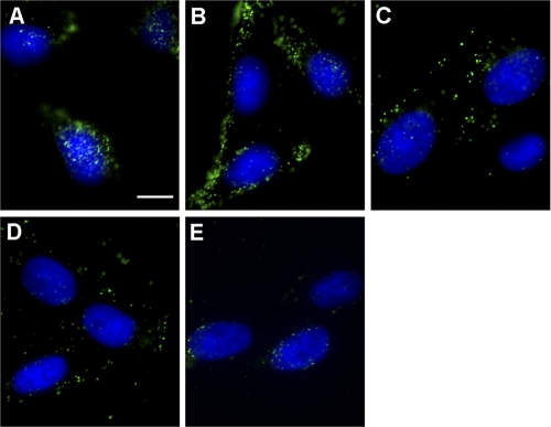
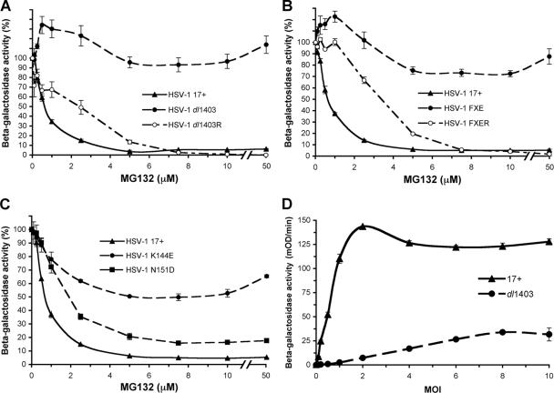
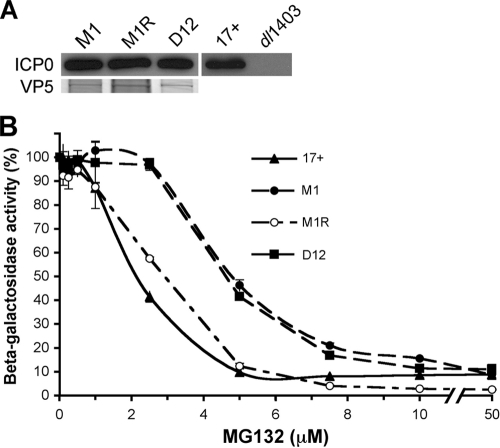
Herpes simplex virus (HSV) entry requires host cell 26S proteasomal degradation activity at a postpenetration step. When expressed in the infected cell, the HSV immediate-early protein ICP0 has E3 ubiquitin ligase activity and interacts with the proteasome. The cell is first exposed to ICP0 during viral entry, since ICP0 is a component of the inner tegument layer of the virion. The function of tegument ICP0 is unknown. Deletion of ICP0 or mutations in the N-terminal RING finger domain of ICP0 results in the absence of ICP0 from the tegument. We show here that these mutations negatively influenced the targeting of incoming capsids to the nucleus. Inhibitors of the chymotrypsin-like activity of the proteasome the blocked entry of virions containing tegument ICP0, including ICP0 mutants that are defective in USP7 binding. However, ICP0-deficient virions were not blocked by proteasomal inhibitors and entered cells via a proteasome-independent mechanism. ICP0 appeared to play a postpenetration role in cells that supported either endocytosis or nonendosomal entry pathways for HSV. The results suggest that ICP0 mutant virions are defective upstream of viral gene expression at a pre-immediate-early step in infection. We propose that proteasome-mediated degradation of a virion or host protein is regulated by ICP0 to allow efficient delivery of entering HSV capsids to the nuclear periphery.


Figures

Fig. 1.
Fig. 1.
Effect of protease inhibitors on HSV entry. (A) CHO-nectin-1 cells were treated with 200 μM E-64d, 100 μM CA074, 50 μM leupeptin, 20 μM TLCK, μM 500 μM pepstatin A, 100 μM phosphoramidon, or 20 μM lactacystin for 15 min at 37°C. HSV-1 KOS-tk12 virus was added at an MOI of 1 for 6 h in the continued presence of inhibitor. (B) Effect of inhibitors of the different catalytic sites of the 20S proteasome. Vero cells were treated with the indicated concentrations of TLCK, YU102, or TPCK for 15 min at 37°C. HSV-1 KOS-tk12 was added for 6 h in the continued presence of inhibitor. The percent beta-galactosidase activity relative to that obtained in the absence of agent is indicated. The data are means of triplicate (A) or quadruplicate (B) determinations with the standard error.
Fig. 2.
Fig. 2.
Subcellular localization of entering virions that lack tegument ICP0. HSV-1 17+ (A), dl1403 (B), FXE (C), K144E (D), or N151D (E) was added to Vero cells in the presence of 0.5 mM cycloheximide. Inocula contained equivalent VP5 units, as determined by protein staining of each virus preparation, corresponding to an MOI of 10 for wild-type 17+. At 2.5 h p.i., the cells were fixed, and capsids were visualized with antibody HA018 to VP5, followed by Alexa 488 secondary antibody. Nuclei were stained with DAPI. For panels A and E, the focus was at the nuclear membrane. For panels B to D, the focus was close to the coverslip. Images are representative of the cell population from at least two independent experiments. Bar, 100 μm.
Fig. 3.
Fig. 3.
Entry of ICP0-null HSV is resistant to inhibition by MG132. (A) Vero cells were treated with MG132 for 15 min at 37°C. Beta-galactosidase-expressing HSV-1 strains KOS-tk12 (wild type) or KOS-7134 (ICP0-deletion) were added (MOI of 1) for 7 h in the continued presence of MG132. (B) MG132 inhibits entry of wild-type HSV strain 17+. CHO-nectin-1 cells were treated with MG132 for 15 min at 37°C. HSV-1 KOS or 17+ was added (MOI of 1) for 6 h in the continued presence of inhibitor. The data are means of quadruplicate determinations with the standard error.
Fig. 4.
Fig. 4.
Effect of MG132 on the entry of ICP0 mutant viruses. (A-C) CHO-nectin-1 cells were treated with MG132 for 15 min at 37°C. HSV-1 17+ wild type, as well as dl1403 or dl1403R (A), FXE or FXER (B), and (C) K144E or N151D, was added (MOI of 1) for 6 h in the continued presence of inhibitor. The activity for each virus in the absence of MG132 was set to 100%. (D) Beta-galactosidase activity expressed by cells infected with HSV-1 wild type or ICP0-null. HSV-1 17+ or dl1403 was added to CHO-nectin-1 cells at the indicated MOI for 6 h. The data are means of quadruplicate determinations with the standard error.
Fig. 5.
Fig. 5.
Inhibition of endosome acidification blocks entry of ICP0-null HSV. CHO-nectin-1 cells were treated with ammonium chloride (NH4Cl) for 15 min at 37°C. HSV-1 17+ (wild type) or dl1403 (ICP0-null) was added (MOI of 1) for 6 h. The percent beta-galactosidase activity relative to that obtained in the absence of NH4Cl is indicated. The data are means of quadruplicate determinations with the standard error.
Fig. 6.
Fig. 6.
Proteasome dependence of entry of HSV with mutations in the USP7 binding domain of ICP0. (A) Association of mutant tegument ICP0 with HSV-1 virions. Extracellular virions of HSV-1 M1, M1R, D12, 17+, or dl1403 were analyzed by SDS-PAGE, followed by Western blotting with antibody 11060 to ICP0. In parallel, VP5 was detected by Coomassie staining to demonstrate equivalent particle loading. (B) Effect of MG132 on the entry of HSV-1 USP7-binding mutants. CHO-nectin-1 cells were treated with MG132 for 15 min at 37°C. The indicated HSV-1 strains were added (MOI of 1) for 6 h, and the beta-galactosidase activity was measured as described in Fig. 4.
Fig. 7.
Fig. 7.
Effect of MG132 on the subcellular localization of entering ICP0 mutant virions. HSV-1 17+ (A), dl1403 (B), FXE (C), K144E (D), or N151D (E) was added to Vero cells in the presence of 25 μM MG132 and 0.5 mM cycloheximide. Inocula contained equivalent VP5 units as determined by protein staining of each virus preparation, corresponding to an MOI of 10 for wild-type 17+. At 2.5 h p.i., cells were fixed, and capsids were visualized with antibody to VP5, followed by Alexa 488 secondary antibody. The nuclei were stained with DAPI. Images are representative of the cell population from at least two independent experiments. Bar, 100 μm.

Similar articles

Cited by

References

    1. Batterson W., Furlong D., Roizman B. 1983. Molecular genetics of herpes simplex virus. VIII. further characterization of a temperature-sensitive mutant defective in release of viral DNA and in other stages of the viral reproductive cycle. J. Virol. 45:397–407 - PMC - PubMed
    1. Boisvert M., Fernandes S., Tijssen P. Multiple pathways involved in porcine parvovirus cellular entry and trafficking toward the nucleus. J. Virol. 84:7782–7792 - PMC - PubMed
    1. Boutell C., Canning M., Orr A., Everett R. D. 2005. Reciprocal activities between herpes simplex virus type 1 regulatory protein ICP0, a ubiquitin E3 ligase, and ubiquitin-specific protease USP7. J. Virol. 79:12342–12354 - PMC - PubMed
    1. Boutell C., Sadis S., Everett R. D. 2002. Herpes simplex virus type 1 immediate-early protein ICP0 and is isolated RING finger domain act as ubiquitin E3 ligases in vitro. J. Virol. 76:841–850 - PMC - PubMed
    1. Brown S. M., Ritchie D. A., Subak-Sharpe J. H. 1973. Genetic studies with herpes simplex virus type 1: the isolation of temperature-sensitive mutants, their arrangement into complementation groups, and recombination analysis leading to a linkage map. J. Gen. Virol. 18:329–346 - PubMed

Publication types

Substances

LinkOut - more resources