. 2011 Jun;85(12):5864-79.
doi: 10.1128/JVI.00155-11. Epub 2011 Apr 6.

Spatial relationships between markers for secretory and endosomal machinery in human cytomegalovirus-infected cells versus those in uninfected cells

Affiliations

Spatial relationships between markers for secretory and endosomal machinery in human cytomegalovirus-infected cells versus those in uninfected cells

Subhendu Das et al. J Virol. 2011 Jun.

Abstract

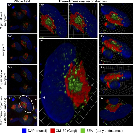
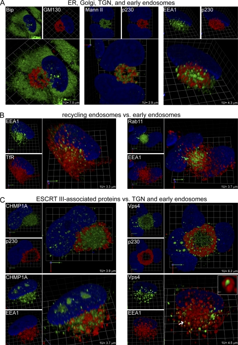
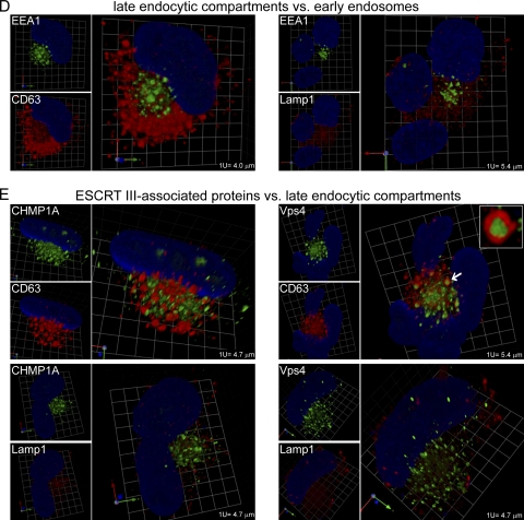
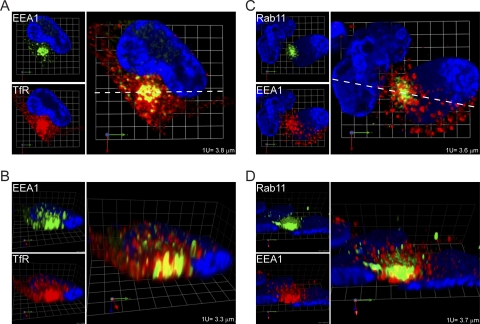
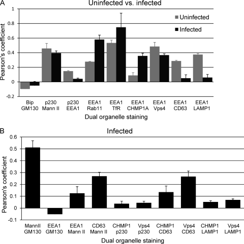
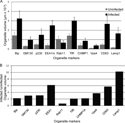
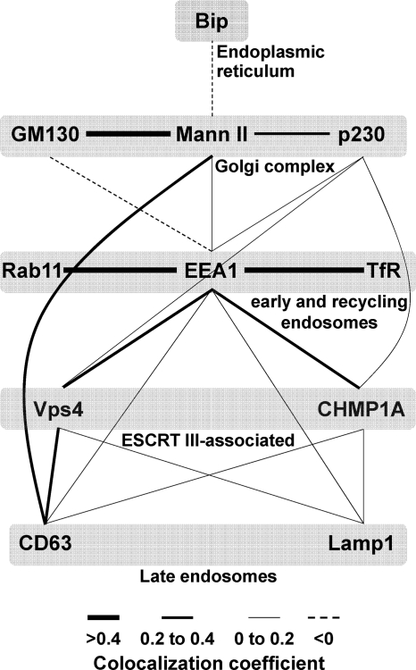
Human cytomegalovirus (HCMV) induces extensive remodeling of the secretory apparatus to form the cytoplasmic virion assembly compartment (cVAC), where virion tegumentation and envelopment take place. We studied the structure of the cVAC by confocal microscopy to assess the three-dimensional distribution of proteins specifically associated with individual secretory organelles. In infected cells, early endosome antigen 1 (EEA1)-positive vesicles are concentrated at the center of the cVAC and, as previously seen, are distinct from structures visualized by markers for the endoplasmic reticulum, Golgi apparatus, and trans-Golgi network (TGN). EEA1-positive vesicles can be strongly associated with markers for recycling endosomes, to a lesser extent with markers associated with components of the endosomal sorting complex required for transport III (ESCRT III) machinery, and then with markers of late endosomes. In comparisons of uninfected and infected cells, we found significant changes in the structural associations and colocalization of organelle markers, as well as in net organelle volumes. These results provide new evidence that the HCMV-induced remodeling of the membrane transport apparatus involves much more than simple relocation and expansion of preexisting structures and are consistent with the hypothesis that the shift in identity of secretory organelles in HCMV-infected cells results in new functional profiles.


Figures

Fig. 1.
Fig. 1.
Relationships between images from single confocal planes and reconstructed 3-D images. HCMV(AD169)-infected lung fibroblasts (HLF cells) were stained for the indicated markers at 120 hpi. Serial 0.5-μm confocal sections were obtained with a Leica TCS SP5 laser-scanning confocal microscope. (A1 to A3) Three of the 23 confocal sections that were used to generate the 3-D reconstruction shown in panel C. The location of each section in the Z-series is indicated. (B) Maximum projection image of the entire field containing the cell from which the 3-D reconstruction was made. This shows that many infected cells have related structures. (C to C7) A collection of views of a 3-D reconstruction of a single binucleated infected cell (circled in panel B). A portion of the nucleus of an overlapping adjacent cell is visible in panels C4 through C7. Panel C7 is a view from the bottom. Grid spacing is 3.31 μm. A video showing the reconstruction rotating in space is available as Video S1 in the supplemental material.
Fig. 2.
Fig. 2.
Relative localization of markers for major secretory organelles, endocytic organelles, and ESCRT III-associated proteins in uninfected lung fibroblasts (HLF cells). (A) Markers of the ER (Bip), Golgi apparatus (GM130 and Mann II), TGN (p230), and early endosomes (EEA1). (B) Markers of early (EEA1) and recycling (TfR and Rab11) endosomes. Structures whose centers are dually positive for EEA1 and TfR and have TfR-rich domains on their surface are indicated with arrows. (C) Markers of ESCRT III-associated proteins (CHMP1A and Vps4). (D) Markers of late endocytic compartments (CD63 and LAMP1). Rabbit polyclonal antibody staining is shown in green, mouse MAb staining in red, and DAPI staining in blue. Grid spacings are indicated for each image. The corresponding videos, Video S2A and S2BCD, are available in the supplemental material.
Fig. 3.
Fig. 3.
Relative localization of markers for major secretory organelles, endocytic organelles, and ESCRT III-associated proteins in HCMV-infected lung fibroblasts (HLF cells). (A) Markers of the ER (Bip), Golgi apparatus (GM130 and Mann II), TGN (p230), and early endosomes (EEA1). (B) Markers of early (EEA1) and recycling (TfR and Rab11) endosomes. (C) Markers of ESCRT III-associated proteins (CHMP1A and Vps4) relative to p230 and EEA1. (D) Markers of late endocytic compartments (CD63 and LAMP1) relative to EEA1. (E) Markers of ESCRT III-associated proteins (CHMP1A and Vps4) versus markers of late endocytic compartments (CD63 and LAMP1). Enlargements of examples of structures described in the text as having the appearance of a sausage wrapped in a bun are provided as insets in the Vps4/EEA1 (C) and Vps4/CD63 (E) images. The objects that were enlarged are indicated with arrows. Rabbit polyclonal antibody staining is shown in green, mouse MAb staining in red, and DAPI staining in blue. Grid spacings are indicated for each image. Corresponding videos, Videos S3A, S3BC, and S3DE, are available in the supplemental material.
Fig. 3.
Fig. 3.
Relative localization of markers for major secretory organelles, endocytic organelles, and ESCRT III-associated proteins in HCMV-infected lung fibroblasts (HLF cells). (A) Markers of the ER (Bip), Golgi apparatus (GM130 and Mann II), TGN (p230), and early endosomes (EEA1). (B) Markers of early (EEA1) and recycling (TfR and Rab11) endosomes. (C) Markers of ESCRT III-associated proteins (CHMP1A and Vps4) relative to p230 and EEA1. (D) Markers of late endocytic compartments (CD63 and LAMP1) relative to EEA1. (E) Markers of ESCRT III-associated proteins (CHMP1A and Vps4) versus markers of late endocytic compartments (CD63 and LAMP1). Enlargements of examples of structures described in the text as having the appearance of a sausage wrapped in a bun are provided as insets in the Vps4/EEA1 (C) and Vps4/CD63 (E) images. The objects that were enlarged are indicated with arrows. Rabbit polyclonal antibody staining is shown in green, mouse MAb staining in red, and DAPI staining in blue. Grid spacings are indicated for each image. Corresponding videos, Videos S3A, S3BC, and S3DE, are available in the supplemental material.
Fig. 4.
Fig. 4.
Regions of high levels of colocalization of early (EEA1) and recycling (TfR and Rab11) endosome markers at the cVAC center. The images are cross-sections through the cVAC of the 3-D reconstructions shown in Fig. 3B, without modification of color intensities. (A and C) Horizontal cross sections (perpendicular to the z-axis) through the 3-D reconstructions (at confocal section 17 of 31 for panel A and 15 of 28 for panel C); (B and D) vertical cross sections (parallel to the z-axis) taken along the dotted white lines in panels A and C. Grid spacings are indicated.
Fig. 5.
Fig. 5.
Colocalization coefficients for pairs of markers. (A) Uninfected versus infected HLF cells. (B) Infected HLF cells. Pearson's threshold colocalization coefficient (as implemented in the Volocity software package) was determined for the indicated pairs of markers. At least 3 cells were evaluated for each pair of markers. Under the analysis conditions used, a voxel (a pixel in three dimensions) was considered positive if both markers were above a specified threshold (36). Under these conditions, voxels that were low positive for one marker and high positive for the other would be considered positive, as would be voxels low or high for both markers. In some instances, images of pairwise comparisons are not shown in the accompanying figures (e.g., Mann II versus GM130 [data not shown]). Mean values are expressed (±standard errors of the means).
Fig. 6.
Fig. 6.
Organelle volumes in uninfected and infected cells. (A) The “Find Objects” algorithm from the Volocity software package was used to identify objects in three-dimensional reconstructions that correspond to objects visible in images such as those shown in Fig. 2 and 3. The net intracellular volume associated with these objects was obtained by summing the volumes of the positive voxels. For EEA1, results were from staining with the mouse MAb. Mean values are expressed (±standard errors of the means). (B) Ratios of organelle volumes in infected cells compared to those in uninfected cells.
Fig. 7.
Fig. 7.
Colocalization network map of membrane transport machinery markers in HCMV-infected cells. Pairwise colocalization data from Fig. 5 were used to generate a map of the colocalization network among the various markers studied here. Three categories of line widths were used that are proportional to the extent of the observed Pearson's colocalization coefficient (high = >0.4; medium = 0.2 to 0.4; low = 0 to 0.2); negative colocalization values are represented as dashed lines. All pairwise comparisons from Fig. 5 are represented, but all possible pairwise combinations were not tested.

Similar articles

Cited by

References

    1. Ahlqvist J., Mocarski E. 2011. Cytomegalovirus UL103 controls virion and dense body egress. J. Virol. 85:5125–5135 - PMC - PubMed
    1. AuCoin D. P., Smith G. B., Meiering C. D., Mocarski E. S. 2006. Betaherpesvirus-conserved cytomegalovirus tegument protein ppUL32 (pp150) controls cytoplasmic events during virion maturation. J. Virol. 80:8199–8210 - PMC - PubMed
    1. Barr F. A., Short B. 2003. Golgins in the structure and dynamics of the Golgi apparatus. Curr. Opin. Cell Biol. 15:405–413 - PubMed
    1. Buchkovich N. J., Maguire T. G., Alwine J. C. 2010. Role of the endoplasmic reticulum chaperone BiP, SUN domain proteins, and dynein in altering nuclear morphology during human cytomegalovirus infection. J. Virol. 84:7005–7017 - PMC - PubMed
    1. Buchkovich N. J., Maguire T. G., Paton A. W., Paton J. C., Alwine J. C. 2009. The endoplasmic reticulum chaperone BiP/GRP78 is important in the structure and function of the HCMV assembly compartment. J. Virol. 83:11421–11428 - PMC - PubMed

Publication types

MeSH terms

Substances

LinkOut - more resources