. 2011 Jun;10(6):929-37.
doi: 10.1158/1535-7163.MCT-10-1109. Epub 2011 Apr 6.

Overexpression of mitotic centromere-associated Kinesin stimulates microtubule detachment and confers resistance to paclitaxel

Affiliations

Overexpression of mitotic centromere-associated Kinesin stimulates microtubule detachment and confers resistance to paclitaxel

Anutosh Ganguly et al. Mol Cancer Ther. 2011 Jun.

Abstract

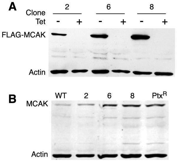
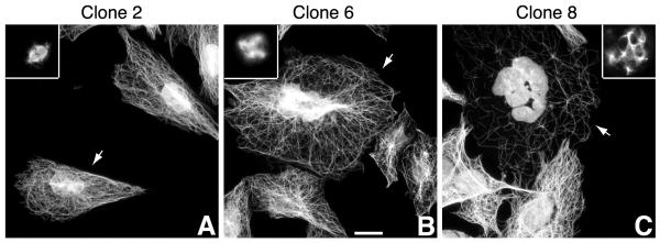
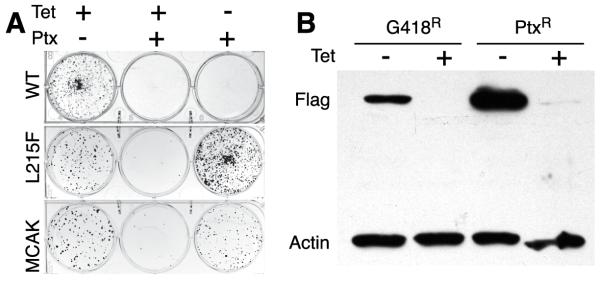
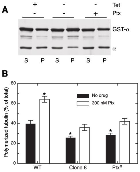
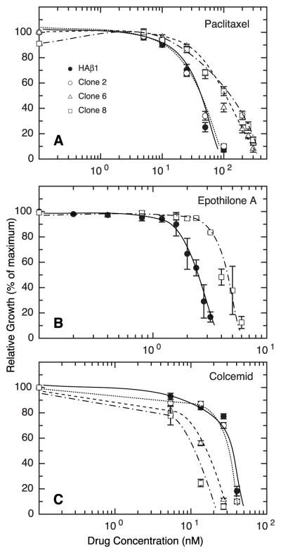
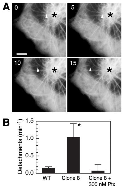
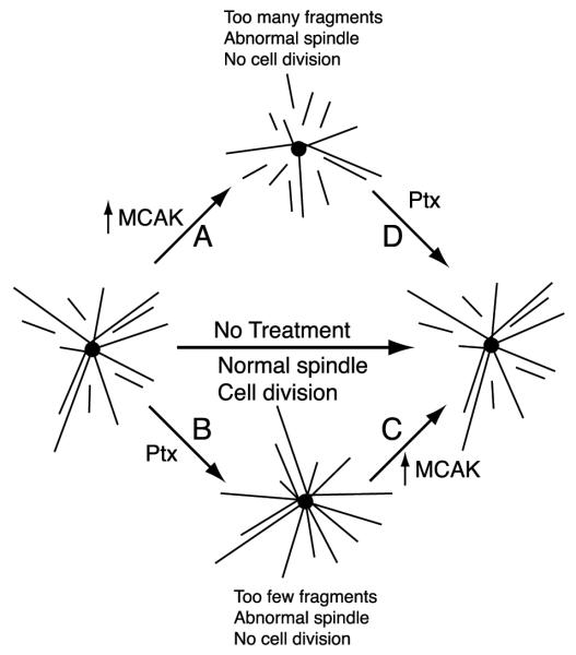
Numerous studies have implicated mutations in tubulin or the overexpression of specific tubulin genes in resistance to microtubule-targeted drugs. Much less is known about the role of accessory proteins that modulate microtubule behavior in the genesis of drug resistance. Here, we examine mitotic centromere-associated kinesin (MCAK), a member of the kinesin family of microtubule motor proteins that has the ability to stimulate microtubule depolymerization, and show that overexpressing the protein confers resistance to paclitaxel and epothilone A, but increases sensitivity to colcemid. Cells transfected with FLAG-tagged MCAK cDNA using a tet-off-regulated expression system had a disrupted microtubule cytoskeleton and were able to survive a toxic concentration of paclitaxel in the absence, but not in the presence of tetracycline, showing that drug resistance was caused by ectopic MCAK production. Moreover, a population that was heterogeneous with respect to FLAG-MCAK expression became enriched with cells that produced the ectopic protein when it was placed under paclitaxel selection. Similar to previously isolated mutants with altered tubulin, paclitaxel resistant cells resulting from MCAK overexpression were found to have decreased microtubule polymer and a seven-fold increase in the frequency of microtubule detachment from centrosomes. These data are consistent with a model for paclitaxel resistance that is based on stability of the attachment of microtubules to their nucleating centers, and they implicate MCAK in the mechanism of microtubule detachment.


Figures

Figure 1
Figure 1
Quantification of MCAK in cell lines stably transfected with FLAG-MCAK. A, CHO cells were transfected with FLAG-MCAK cDNA and stable cell lines were selected in G418. Western blots of 3 clones grown in the presence (+) and absence (−) of tetracycline (Tet) for 24 h are shown. An antibody specific for the FLAG tag was used. An antibody to actin was also included as a loading control. B, similar cell lysates were compared on western blots using an antibody that recognizes endogenous as well as transfected MCAK. The gel included non-transfected wild-type CHO cells (WT) and FLAG-MCAK transfected cells selected in paclitaxel (Ptx) as controls.
Figure 2
Figure 2
Immunofluorescence of cell lines expressing FLAG-MCAK. Stable clones expressing FLAG-MCAK were mixed with non-transfected wild-type cells for an internal control, grown for 2 d, and stained with antibodies to tubulin and FLAG tag (not shown). DNA was stained with DAPI to label nuclei. Arrows point to the cells that express FLAG-MCAK. Insets show typical mitotic cells. Scale bar, 10 μm.
Figure 3
Figure 3
MCAK overexpression confers paclitaxel resistance. Non-transfected wild-type (WT) CHO cells, CHO cells transfected with HAβ1-tubulin containing an L215F mutation, and a G418 selected population of CHO cells transfected with FLAG-MCAK were seeded into dishes in the presence (+) or absence (−) of tetracycline (Tet) and/or paclitaxel (Ptx). The cells were incubated for 7 d until colonies appeared and the dishes were stained with methylene blue and photographed. Cells in the first column were seeded at low density to estimate the relative number of viable cells for each cell line. Cells in the following 2 columns were under selective pressure from 300 nM paclitaxel and were therefore seeded at a 50-fold higher density. (B) CHO cells transfected with FLAG-MCAK were selected in G418 to obtain a stable cell population. These cells were reselected in 300 nM paclitaxel (Ptx) to obtain a drug-resistant population. Both cell populations were grown with (+) and without (−) tetracycline (Tet) for 24 h and then analyzed by western blots with an antibody to the FLAG tag and an antibody to actin as a gel loading control.
Figure 4
Figure 4
Effect of FLAG-MCAK expression on microtubule content. A, Clone 8 cells were grown 24 h in the presence (+) or absence (−) of tetracycline (Tet) or paclitaxel (Ptx), and cell lysates were centrifuged to separate microtubules in the pellet fraction (P) from soluble tubulin in the supernatant (S). GST-α-tubulin was added to both fractions to act as an internal control for possible losses of material during subsequent steps. The western blot was stained with an antibody specific for α-tubulin. Note that there is less tubulin in the pellet fraction when the cells were grown without tetracycline to induce FLAG-MCAK expression and that the tubulin level returned to near normal when paclitaxel was present during tetracycline withdrawal. Minor bands are degradation products of GST-α-tubulin. B, wild-type (WT), Clone 8, and transfected cells selected for resistance to paclitaxel (PtxR) were incubated overnight in the presence or absence of 300 nM paclitaxel and their microtubule polymer was separated from soluble tubulin by centrifugation. Tubulin in the supernatant and pellet fractions was normalized to the amount of GST-α, and the extent of polymerization was calculated as the amount in the pellet (P) divided by the total tubulin (S + P). Bars represent the mean ± SD from 3 experiments run in triplicate. *p < 0.01 relative to WT without drug.
Figure 5
Figure 5
Drug resistance of cells expressing FLAG-MCAK. Clones 2, 6, and 8 which produce increasing amounts of FLAG-MCAK were compared to cells stably transfected with wild-type HAβ1-tubulin for their sensitivity to paclitaxel (A), epothilone A (B), and colcemid (C). The cells were seeded at low density and incubated 7 d until visible colonies appeared. Total relative cell growth was determined and plotted against the drug concentration. The experiment was run in triplicate and repeated 3 times. Error bars, SD.
Figure 6
Figure 6
Microtubule detachment in MCAK overexpressing cells. A, Clone 8 cells grown in the absence of tetracycline for 2 d were transfected with EGFP-MAP4. Successive images taken 5 s apart are shown. Arrowheads mark the minus end of a microtubule seen to detach from the centrosome (asterisk). Scale bar, 2 μm. B, the frequencies of microtubule detachment in wild-type (WT), Clone 8, and Clone 8 treated with 300 nM paclitaxel (Ptx) were each calculated from 7-19 cells over a total observation time of 25-80 min. The values shown are the mean ± SEM. *p < 0.01.
Figure 7
Figure 7
Model for the role of MCAK in spindle formation and drug resistance. The model is based on the idea that normal spindle assembly and function requires an appropriate mixture of centrosomal microtubules and non-centrosomal fragments that are generated by microtubule detachment from the spindle poles. MCAK and paclitaxel have opposing effects on this process: increased MCAK activity (A) stimulates microtubule detachment leading to more fragments; treatment with paclitaxel (B) suppresses microtubule detachment leading to fewer fragments. Cells that are treated with a toxic concentration of paclitaxel experience problems with spindle function because there are too few fragments, but this condition can be alleviated by increasing MCAK activity (C). Thus, cells with elevated MCAK activity are drug resistant. At very high levels of MCAK activity cells may have spindle defects because there are too many microtubule fragments. Treatment with paclitaxel (D) will reduce the number of fragments and allow the cells to grow normally. Thus cells with very high MCAK activity are predicted to be drug dependent for growth.

Similar articles

Cited by

References

    1. Cabral F. Factors determining cellular mechanisms of resistance to antimitotic drugs. Drug Resistance Updates. 2001;3:1–6. - PubMed
    1. Ems-McClung SC, Walczak CE. Kinesin-13s in mitosis: Key players in the spatial and temporal organization of spindle microtubules. Semin Cell Dev Biol. 2010;21:276–82. - PMC - PubMed
    1. Lawrence CJ, Dawe RK, Christie KR, Cleveland DW, Dawson SC, Endow SA, et al. A standardized kinesin nomenclature. J Cell Biol. 2004;167:19–22. - PMC - PubMed
    1. Helenius J, Brouhard G, Kalaidzidis Y, Diez S, Howard J. The depolymerizing kinesin MCAK uses lattice diffusion to rapidly target microtubule ends. Nature. 2006;441:115–9. - PubMed
    1. Ogawa T, Nitta R, Okada Y, Hirokawa N. A common mechanism for microtubule destabilizers-M type kinesins stabilize curling of the protofilament using the class-specific neck and loops. Cell. 2004;116:591–602. - PubMed

Publication types

MeSH terms