. 2011 May;9(5):589-602.
doi: 10.1158/1541-7786.MCR-10-0565. Epub 2011 Apr 6.

Differential roles for the interferon-inducible IFI16 and AIM2 innate immune sensors for cytosolic DNA in cellular senescence of human fibroblasts

Affiliations

Differential roles for the interferon-inducible IFI16 and AIM2 innate immune sensors for cytosolic DNA in cellular senescence of human fibroblasts

Xin Duan et al. Mol Cancer Res. 2011 May.

Abstract

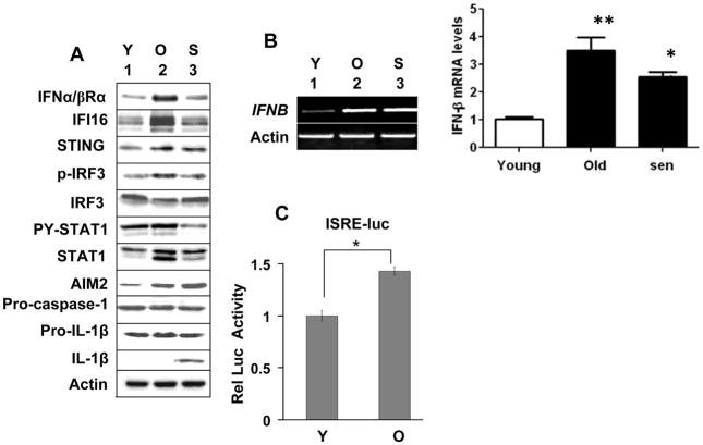
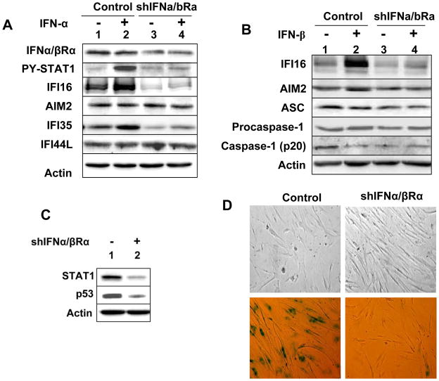
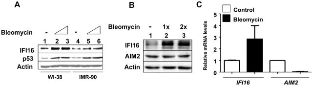
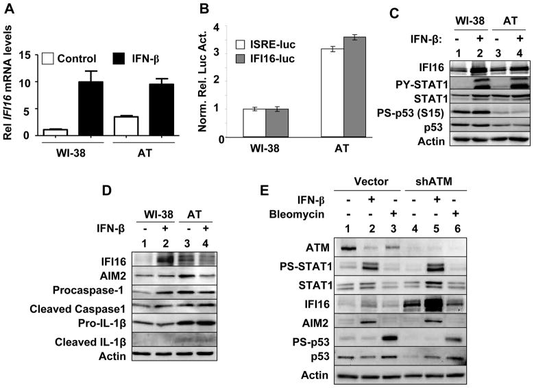
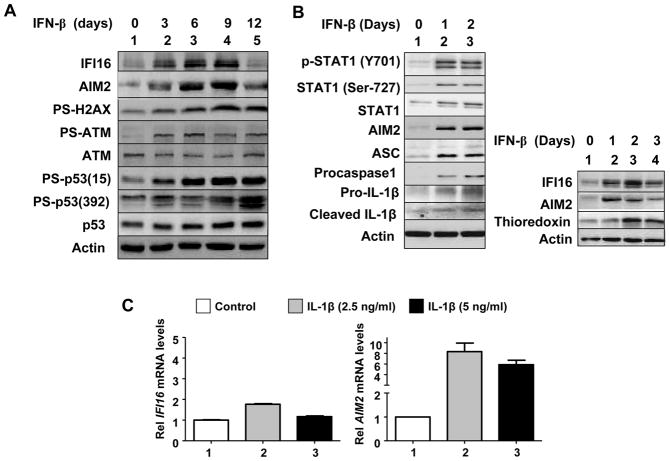
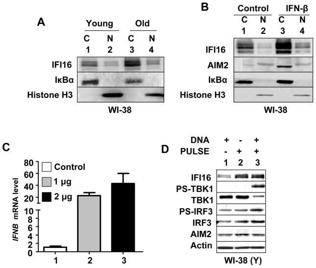
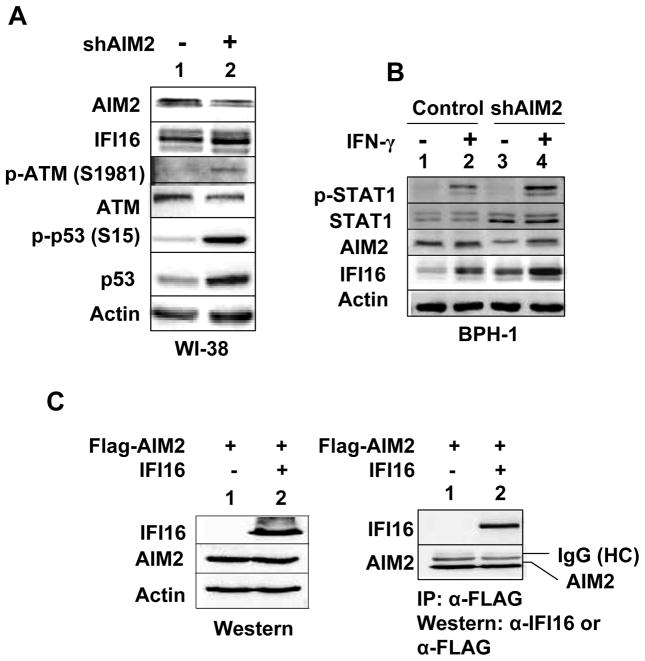
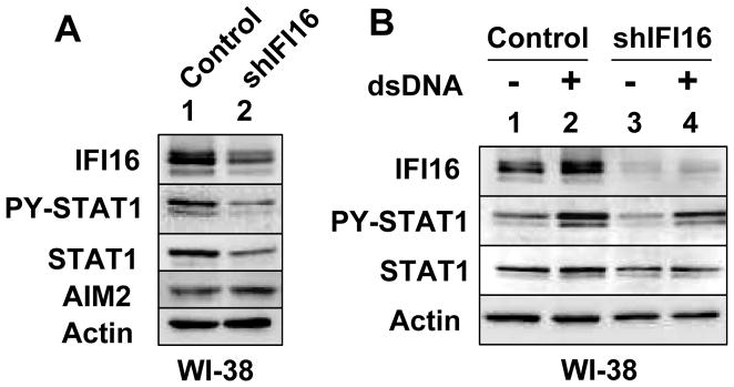
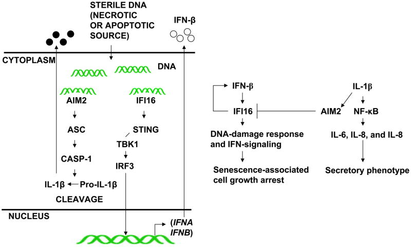
The IFN-inducible IFI16 and AIM2 proteins act as innate immune sensors for cytosolic double-stranded DNA (dsDNA). On sensing dsDNA, the IFI16 protein induces the expression of IFN-β whereas the AIM2 protein forms an inflammasome, which promotes the secretion of IL-1β. Given that the knockdown of IFI16 expression in human diploid fibroblasts (HDF) delays the onset of cellular senescence, we investigated the potential roles for the IFI16 and AIM2 proteins in cellular senescence. We found that increased IFI16 protein levels in old (vs. young) HDFs were associated with the induction of IFN-β. In contrast, increased levels of the AIM2 protein in the senescent (vs. old) HDFs were associated with increased production of IL-1β. The knockdown of type I IFN-α receptor subunit, which reduced the basal levels of the IFI16 but not of the AIM2, protein delayed the onset of cellular senescence. Accordingly, increased constitutive levels of IFI16 and AIM2 proteins in ataxia telangiectasia mutated (ATM) HDFs were associated with the activation of the IFN signaling and increased levels of IL-1β. The IFN-β treatment of the young HDFs, which induced the expression of IFI16 and AIM2 proteins, activated a DNA damage response and also increased basal levels of IL-1β. Interestingly, the knockdown of AIM2 expression in HDFs increased the basal levels of IFI16 protein and activated the IFN signaling. In contrast, the knockdown of the IFI16 expression in HDFs decreased the basal and dsDNA-induced activation of the IFN signaling. Collectively, our observations show differential roles for the IFI16 and AIM2 proteins in cellular senescence and associated secretory phenotype.


Conflict of interest statement

Disclosure of Potential Conflicts of Interest

The authors have no financial conflicts of interest.

Figures

FIGURE 1
FIGURE 1
Differential expression and roles for the IFI16 and AIM2 proteins during cellular senescence of HDFs. A, Total cell extracts prepared from young (Y), old (O), or senescent (S) WI-38 HDFs were analyzed by immunoblotting using antibodies specific to the indicated proteins. B, Total RNA isolated from young (Y), old (O), or senescent (S) WI-38 HDFs were analyzed by semi-quantitative PCR (left panel) or quantitative real-time PCR (right panel) for the indicated genes. The ratio of the test gene to actin mRNA was calculated in units (one unit being the ratio of the test gene to actin mRNA). The relative steady-state levels of IFNB mRNA in young HDFs are indicated as 1. Results are mean values of triplicate experiments and error bars represent standard deviation (*p <0.05; ** p <0.01). C, Sub-confluent cultures of young (Y) or old (O) WI-38 HDFs were infected with Cignal Lenti ISRE Reporter (Luc) virus as suggested by the supplier. The infected cells were harvested after 40-44 h to assays for the firefly and Renilla luciferase activitiesas described in methods.Normalized relative firefly luciferase activity is shown.
FIGURE 2
FIGURE 2
The knockdown of the IFNα/βRα subunit expression in HDFs decreases IFI16 expression and delays the onset of a senescence phenotype. A, Sub-confluent cultures of young WI-38 HDFs infected with control lentivirus (control) or virus encoding for shIFNa/bRa RNA (shIFNa/bRa) were either left untreated or treated with IFN-α for 18 h. After the treatment, total cell extracts were prepared and analyzed by immunoblotting using antibodies specific to the indicated proteins. B, Control or shIFNa/bRa HDFs described in panel A were either left untreated or treated with IFN-β for 18 h. Total cell extracts were analyzed by immunoblotting. C, Total cell extracts from control or shIFNa/bRa HDFs were analyzed by immunoblotting for the indicated proteins. D, Phase contrast photographs indicating morphological changes (top two panels) and differences in the number of SA-β-gal-positive cells (bottom two panels) between control HDFs and HDFs after the knockdown of the IFNa/bRa subunit expression.
FIGURE 3
FIGURE 3
DNA-damage response also contributes to the constitutive levels of the IFI16 protein. A, Sub-confluent cultures of young WI-38 or IMR-90 HDFs were either left untreated (lanes 1 and 4) or treated with increasing units of bleomycin (lanes 2, 3, 5 and 6) for 24 h. After the treatment, total cell extracts were analyzed by immunoblotting for the indicated proteins. B, Sub-confluent cultures of young WI-38 were either left untreated (lanes 1) or treated with increasing units of bleomycin (lanes 2 and 3) for 24 h. After the treatment, total cell extracts were analyzed by immunoblotting for the indicated proteins. C, Sub-confluent cultures of young WI-38 were either left untreated or treated with bleomycin for 24 h. After the treatment, total RNA was analyzed by quantitative real-time PCR for IFI16 and AIM2 mRNA levels. The ratio between the test gene (the IFI16 or AIM2) mRNA levels to actin mRNA was calculated in units (one unit being the ratio of the test gene to actin mRNA). The relative steady-state levels of IFI16 mRNA in control HDFs are indicated as 1.
FIGURE 4
FIGURE 4
Constitutively increased levels of the IFI16 protein in ataxia telangiectasia (AT) HDFs are associated with the activation of IFN-signaling. A, Sub-confluent cultures of young WI-38 or AT HDFs were either left untreated or treated with IFN-β for 18 h. After the treatment, total RNA was extracted and analyzed by quantitative real-time PCR for steady-state levels of IFI16 mRNA. The ratio between the test gene the IFI16 mRNA levels to actin mRNA was calculated in units (one unit being the ratio of the test gene to actin mRNA). The relative steady-state levels of IFI16 mRNA in WI-38 control HDFs are indicated as 1. B, Sub-confluent culture of young WI-38 or AT HDFs was transfected with infected with either ISRE-luc or IFI-16-luc reporter plasmid (1.8 μg) along with a second pRL-TK reporter (0.2 μg). The transfected cells were harvested after 40-44 h to assays for the firefly and Renilla luciferase activitiesas described in methods.Normalized relative firefly luciferase activity in WI-38 cells is shown as 1. C, Sub-confluent cultures of young WI-38 or AT HDFs were either left untreated (lanes 1 and 3) or treated with IFN-β (1000 u/ml) for 18 h. After the treatment, total cell extracts were analyzed by immunoblotting for the indicated proteins. D, Sub-confluent cultures of young WI-38 or AT HDFs were either left untreated (lanes 1 and 3) or treated with IFN-β (1000 u/ml) for 18 h. After the treatment, total cell extracts were analyzed by immunoblotting for the indicated proteins. E, Sub-confluent cultures of young WI-38 HDFs that were infected with control virus (Vector) or shATM virus (shATM) were left untreated (lanes 1 and 4), treated with IFN-β (lanes 2 and 5), or bleomycin (lanes 3 and 6) for 18 h. After the treatment, total cell extracts were analyzed by immunoblotting for the indicated proteins.
FIGURE 5
FIGURE 5
Prolonged IFN-β treatment of young HDFs increases levels of IFI16, AIM2, and proinflammatory cytokine IL-1β. A and B, For treatment of young WI-38 HDFs with IFN-β, cultures at ~20% confluence were treated with IFN-β for the indicated days (medium was changed after every two days and fresh IFN-β was added). After the treatment, HDFs were harvested and total cell lysates were analyzed by immunoblotting for the levels of the indicated proteins. C, Cultures of young WI-38 were either left untreated or treated with the indicated amount of the human recombinant IL-β for 18 h. After the treatment, total RNA was extracted and analyzed by quantitative real-time PCR for steady-state levels of IFI16 (left panel) or AIM2 (right panel) mRNA. The ratio between the test gene (the IFI16 or AIM2) mRNA levels to actin mRNA was calculated in units (one unit being the ratio of the test gene to actin mRNA). The relative steady-state levels of IFI16 or AIM2 mRNA in WI-38 control HDFs are indicated as 1.
FIGURE 6
FIGURE 6
IFI16 protein is primarily detected in the cytoplasm of old or IFN-β treated HDFs and nucleofection of young HDFs with dsDNA induces the IFN-β expression. A, Cultures of young (PD ~30) proliferating or old (PD ~50) WI-38 HDFs were harvested and cells were subjected to the nuclear and cytoplasmic fractionation. The fractions containing equal amounts of proteins were analyzed by immunoblotting using antibodies specific to the indicated proteins. B, Cultures of young proliferating WI-38 HDFs were either left untreated or treated with IFN-β for 24 h. After the treatment, cells were subjected to the nuclear and cytoplasmic fractionation. The fractions containing approximately equal amounts of proteins were analyzed by immunoblotting. C, Young proliferating (PD ~30) WI-38 HDFs were nucleofected without DNA (control) or with the indicated amounts of plasmid DNA (GFP plasmid) as described in methods. After 18 h of nucleofection, total RNA was extracted and analyzed by quantitative real-time PCR for steady-state levels of IFNB mRNA. The ratio between the IFNB mRNA levels to actin mRNA was calculated in units (one unit being the ratio of the IFNB mRNA to actin mRNA). The relative steady-state levels of IFNB mRNA in control HDFs are indicated as 1. D, Young proliferating WI-38 HDFs were either left without nucleofection or nucleofected without DNA or nucleofected with 2 μg of plasmid DNA. After 18 h of nucleofection, total cell lysates were analyzed by immunoblotting for the indicated proteins.
FIGURE 7
FIGURE 7
Knockdown of the AIM2 expression in cells increases the IFI16 protein levels. A, Total cell extracts prepared from WI-38 HDFs that were infected with control virus or shAIM2 virus were analyzed by immunoblotting for the indicated proteins. B, Sub-confluent cultures of the human benign prostate hyperplasia-1 (BPH-1) that were infected with control virus (control) or shAIM2 virus (shATM) were either left untreated (lanes 1 and 3) or treated with IFN-γ (lanes 2 and 4) for 18 h. After the treatment, total cell extracts were analyzed by immunoblotting for the indicated proteins. C, Cultures of human embryonic kidney cells (HEK-293) were either transfected with pCMV-AIM2 plasmid (2 μg) allowing the expression of the FLAG-tagged human AIM2 protein or along with pCMV-IFI16 plasmid allowing the expression of IFI16 protein. 40 h after the transfections, total cell lysates were analyzed by immunoblotting using antibodies specific to the IFI16 protein or FLAG-tag (left panel). Total cell extracts were also subjected to immunoprecipitation using antibodies to FLAG-tag and immunoprecipitates were analyzed by immunoblotting (right panel).
FIGURE 8
FIGURE 8
Knockdown of IFI16 expression in HDFs reduces the constitutive and dsDNA-induced activation of IFN-signaling. A, Total cell extracts prepared from the young WI-38 HDFs that were infected with control virus (Vector) or shIFI16 virus (shIFI16) were analyzed by immunoblotting for the indicated proteins. B, Young WI-38 HDFs that were infected with control virus (Vector) or shIFI16 virus (shIFI16) were either nucleofected without (lanes 1 and 3) or with (lanes 2 and 4) dsDNA. 18 h after nucleofections, cells were harvested and total cell extracts were analyzed by immunoblotting for the indicated proteins.
FIGURE 9
FIGURE 9
Proposed roles of the IFI16 and AIM2 proteins in cellular senescence-associated cell growth arrest and secretory phenotype in HDFs.

References

    1. Campisi J. Cellular senescence as a tumor-suppressor mechanism. Trends Cell Biol. 2001;11:S27– 31. - PubMed
    1. Campisi J. Cancer and ageing: rival demons? Nat Rev Cancer. 2003;3:339–49. - PubMed
    1. Young AR, Narita M. SASP reflects senescence. EMBO Rep. 2009;10:228–30. - PMC - PubMed
    1. Mammone T, Gan D, Foyouzi-Youssefi R. Apoptotic cell death increases with senescence in normal human dermal fibroblast cultures. Cell Biol Int. 2006;30:903–9. - PubMed
    1. Lehmann BD, Paine MS, Brooks AM, McCubrey JA, Renegar RH, et al. Senescence-associated exosome release from human prostate cancer cells. Cancer Res. 2008;68:7864–71. - PMC - PubMed

Publication types

MeSH terms