. 2011 Apr 6;31(14):5262-70.
doi: 10.1523/JNEUROSCI.4897-10.2011.

Versican V0 and V1 direct the growth of peripheral axons in the developing chick hindlimb

Affiliations

Versican V0 and V1 direct the growth of peripheral axons in the developing chick hindlimb

Shilpee Dutt et al. J Neurosci. .

Abstract

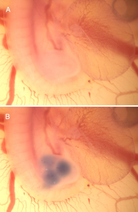
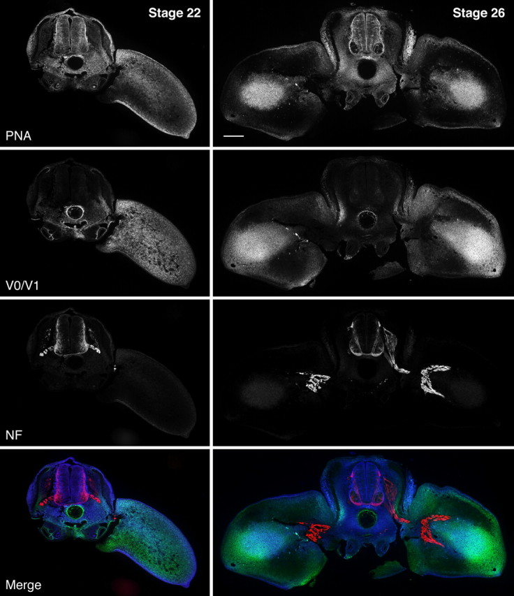
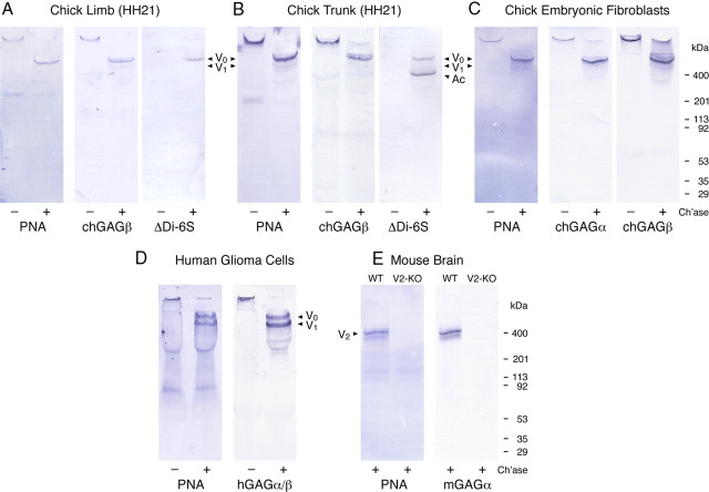
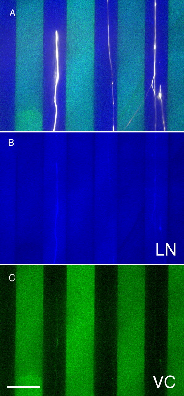
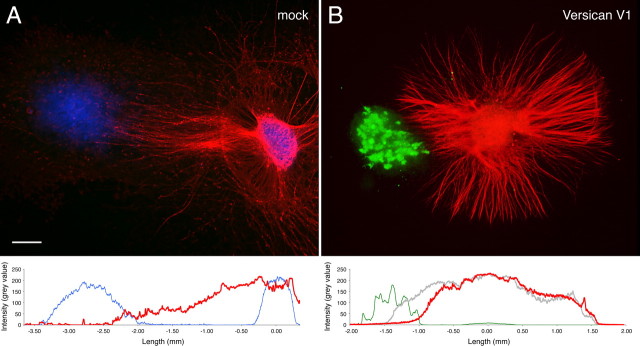
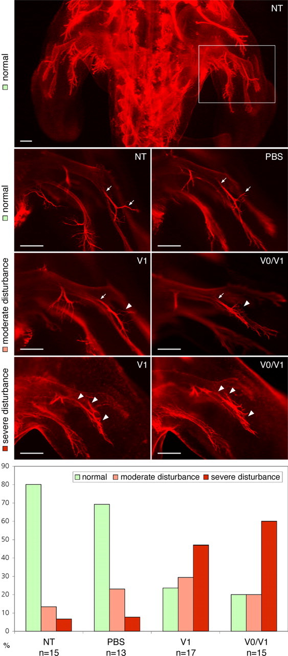
Peanut agglutinin-binding disaccharides and chondroitin sulfate mark transient mesenchymal barriers to advancing motor and sensory axons innervating the hindlimbs during chick development. Here we show that the vast majority of these carbohydrates are at the critical stage and location attached to the versican splice variants V0 and V1. We reveal that the isolated isoforms of this extracellular matrix proteoglycan suppress axon extension at low concentrations and induce growth cone collapse and rapid retraction at higher levels. Moreover, we demonstrate that versican V0 and/or V1, recombinantly expressed in collagen-I gels or ectopically deposited in the hindlimbs of chicken embryos in ovo, cause untimely defasciculation and axon stalling. Consequently, severe disturbances of nerve patterning are observed in the versican-treated embryos. Our experiments emphasize the inhibitory capacity of versicans V0 and V1 in axonal growth and evidence for their function as basic guidance cues during development of the peripheral nervous system.


Figures

Figure 1.
Figure 1.
Chick embryo at HH21/HH22 before (A) and after (B) in ovo injection of the trypan blue-containing versican solution.
Figure 2.
Figure 2.
Temporospatial relationship of PNA-binding sites, versican V0/V1, and axonal growth in the developing hindlimb. Triple lectin/immunofluorescence stainings of cross-sections of HH22 and HH26 chicken embryos at hindlimb level reveal the primarily overlapping patterns of PNA ligands (dark blue in merged image) and versican V0/V1 (green). At HH22, both components are ubiquitously distributed in the perinotochordal region and the early limb bud but absent from the pathways of motor and sensory axons (red), which have reached the plexus region and are momentarily hindered from entering the limb. At HH26, versican and PNA-binding sites have disappeared from the prospective axon routes but persist in the pelvic girdle precursor, in the forming dermis, and the condensing mesenchyme adumbrating the future leg bones. Scale bar, 200 μm.
Figure 3.
Figure 3.
Identification of versicans as major PNA-binding proteins. Extracts of hindlimbs (A) and trunks (B) from HH21 chick embryos, supernatants of chick embryonic fibroblasts (C), and of human U251 glioma cell cultures (D) and brain extracts of P30 wild-type (WT) and versican V0/V2 knock-out mice (E; KO) were analyzed by lectin and immunoblotting with PNA and antibodies against versicans and against chondroitin-6-sulfate stubs (ΔDi-6S), respectively. Some of the samples were digested with chondroitinase ABC (+) to remove the GAG side chains. The domain-specific antibodies against versican are reactive with the chick variants V0/V1 (chGAGβ), the chick and mouse isoforms V0/V2 (chGAGα, mGAGα), or all proteoglycan variants of human versican (hGAGα/β). The bands of the different core glycoproteins of versicans (V0, V1, and V2) and of aggrecan (Ac) are indicated by arrowheads.
Figure 4.
Figure 4.
Versican overrides the neurite growth-promoting activity of laminin-1 in a stripe-choice assay. A, Overlay of false color images of a triple immunofluorescence staining of laminin-1 (LN; blue), versican (VC; green), and neurofilaments (white). Despite the rather uniform distribution of the growth-promoting substrate laminin-1, neurites from E10 chick dorsal root ganglions avoid stripes coated with laminin-1 plus versican V0/V1 (100 μg/ml coating concentration) and advance on lanes coated with laminin-1 alone. The laminin- and the versican-specific stainings are also depicted separately in B and C, respectively. Scale bar, 50 μm.
Figure 5.
Figure 5.
Versican-mediated inhibition of neurite outgrowth on fibronectin. Although neurites from an E10 chick dorsal root ganglion extend freely on alternating substrate stripes coated with 20 and 100 μg/ml fibronectin (A), they are rapidly forced into versican-free lanes on alternating stripes of fibronectin alone (coated with 20 μg/ml) and fibronectin plus versican V0/V1 (100 μg/ml each) substrates (B). Phase-contrast and versican-specific immunofluorescence images. Scale bar, 100 μm.
Figure 6.
Figure 6.
Inhibitory potential of versican V0/V1 on neurite outgrowth in stripe-choice assays. Neurites from E10 chick dorsal root ganglions grow on laminin-1 stripes but avoid alternating lanes coated with laminin-1 plus intact or core glycoprotein preparations of versican V1 (B, C and G, H, respectively) or of a V0/V1 mix (D, E and I, J). For the coating of the test stripes, a 100 μg/ml laminin-1 solution was supplemented with variable amounts of versican. The corresponding versican concentrations are indicated above the panels. On the versican-free control lanes, 20 μg/ml laminin-1 was applied. The stripe patterns of the control assays (A, F) were prepared analogously, but versican was omitted in the test stripes; control (F) included in addition chondroitinase and enzyme buffer in the test lanes. Neurofilaments (red) and versican (green) have been visualized by double immunofluorescence staining. Ratios (±SD) of averaged gray values of the axon-specific red fluorescence in versican-free versus the adjacent versican-containing lanes are shown below the panels. Scale bar, 100 μm.
Figure 7.
Figure 7.
Growth cone collapse and retraction of DRG neurites after contact with versican-containing substrate. Still images from time-lapse video microscopy (A–F) documenting the axon growth behavior of E10 chick DRG axons on a laminin-1 (LN) substrate either containing or lacking versican V0/V1 (VC). The boundary to the area coated with laminin plus versican V0/V1 (100 μg/ml each) is marked with a stippled line. At 0 and 63 min, the growth cone (arrowhead) is moving forward in direction of the versican-containing region. Contacting versican after 69 min, it collapses and the axon rapidly retracts back toward the cell body. After 87 min, the original position is reached (F). Scale bar: A–F, 25 μm. Immunofluorescence overlays of substrate stainings of versican (G, H, J–L) or laminin-1 (I) and axonin-1-specific neurite-labeling reveal growth cones with partly to fully extended structures during advancement on a versican-free zone between versican-containing stripes (G, H) or across the border of two differentially coated laminin-1 control areas (I; 100 μg/ml coating above and 20 μg/ml below arrowhead). A fully collapsed morphology is, however, invariably observed near the ends of versican-free lanes (arrowheads in J, K). L, A neuronal cell body near the versican-containing surface projects a long neurite perpendicularly away from versican, whereas a short second process extends strictly parallel to the border. Scale bar: G–L, 10 μm.
Figure 8.
Figure 8.
Inhibition of axonal outgrowth by versican V1-expressing COS aggregates in collagen-I gel. Neurites from E10 chick DRG extend radially within the collagen gel but defasciculate and stop near the COS cell aggregate recombinantly expressing versican V1. No such effect is observed in the control experiment with a COS cell aggregate transfected with the empty vector. Neurofilaments (red) and versican (green) are visualized by double immunofluorescence staining. Hoechst nuclear stain (blue) reveals the cellular localizations in the control. Fluorescence profiles averaged over a bandwidth of 200 μm reveal the decline of neurite density (red line in B) already 0.5 mm away from the versican-expressing (green line) COS cells in relation to the profile of a nearby control sector, in which neurites pass by the aggregate unhindered (gray line). Profiles of neurites and COS aggregates are virtually non-overlapping contrasting the control experiment (A; Hoechst, blue line; neurites, red line). Scale bar, 400 μm.
Figure 9.
Figure 9.
Consequences of ectopic deposition of versicans in developing chick hindlimb in ovo. Whole-mount immunofluorescence stainings of neurofilaments in HH25 embryos injected beforehand with PBS, versican V0/V1, or versican V1 preparations into the hindlimb are compared with nontreated controls (NT). The ectopic deposition of versican V0/V1 and V1 interferes with the normal growth, fasciculation, and branching of secondary nerve fibers emerging from dorsal trunk of the sciatic plexus. Normal branch points are indicated with arrows and aberrations with arrowheads. Scale bars, 200 μm. The proportions of normal, moderately, and severely disturbed growth patterns were recorded in a blind study revealing the clear effect of the versican injections.

References

    1. Bandtlow CE, Zimmermann DR. Proteoglycans in the developing brain - new conceptual insights for old proteins. Physiol Rev. 2000;80:1267–1290. - PubMed
    1. Davies JA, Cook GM, Stern CD, Keynes RJ. Isolation from chick somites of a glycoprotein fraction that causes collapse of dorsal root ganglion growth cones. Neuron. 1990;4:11–20. - PubMed
    1. Domowicz M, Li H, Hennig A, Henry J, Vertel BM, Schwartz NB. The biochemically and immunologically distinct CSPG of notochord is a product of the aggrecan gene. Dev Biol. 1995;171:655–664. - PubMed
    1. Dours-Zimmermann MT, Maurer K, Rauch U, Stoffel W, Fässler R, Zimmermann DR. Versican V2 assembles the extracellular matrix surrounding the nodes of ranvier in the CNS. J Neurosci. 2009;29:7731–7742. - PMC - PubMed
    1. Dutt S, Kléber M, Matasci M, Sommer L, Zimmermann DR. Versican V0 and V1 guide migratory neural crest cells. J Biol Chem. 2006;281:12123–12131. - PubMed

Publication types

MeSH terms

LinkOut - more resources