. 2011 Apr 6;31(14):5325-34.
doi: 10.1523/JNEUROSCI.6156-10.2011.

The basis for diminished functional recovery after delayed peripheral nerve repair

Affiliations

The basis for diminished functional recovery after delayed peripheral nerve repair

Tessa Gordon et al. J Neurosci. .

Abstract

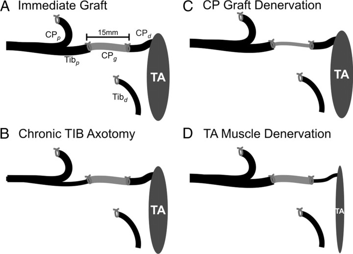
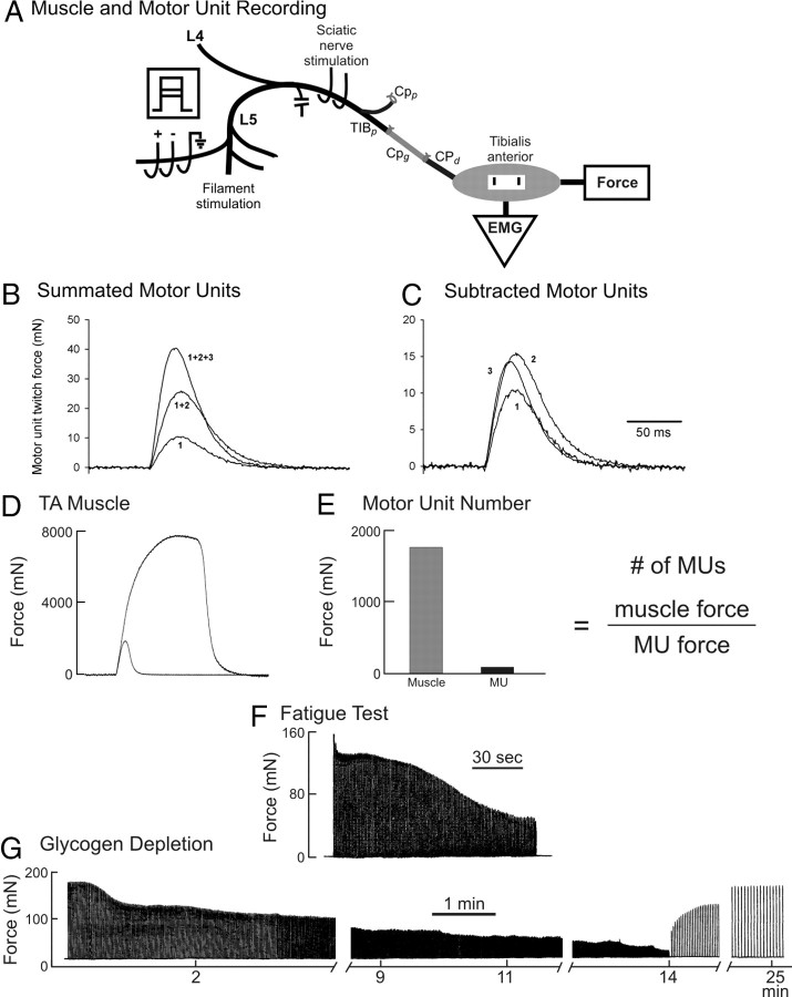
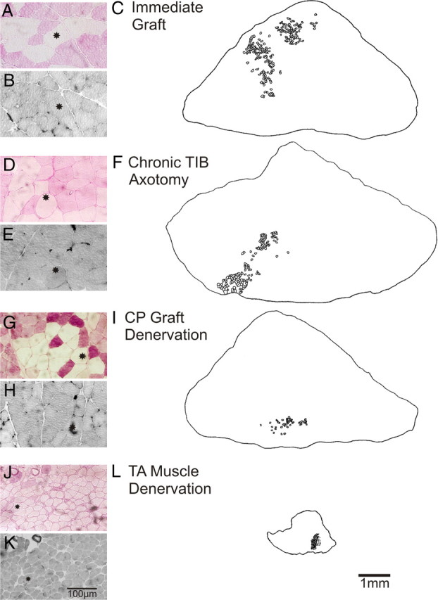
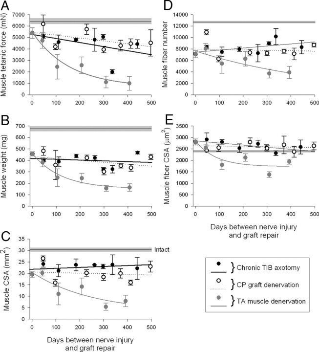
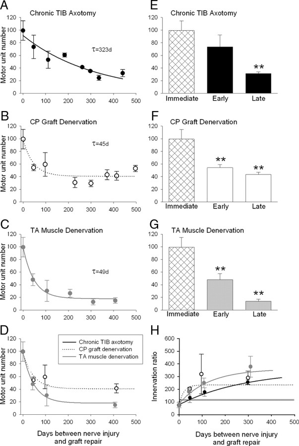
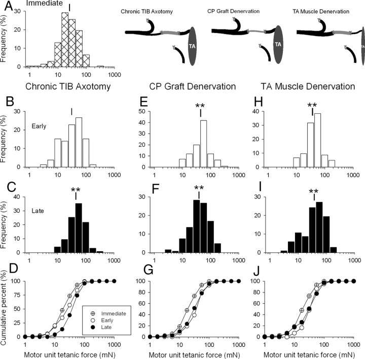
The postsurgical period during which neurons remain without target connections (chronic axotomy) and distal nerve stumps and target muscles are denervated (chronic denervation) deleteriously affects functional recovery. An autologous nerve graft and cross-suture paradigm in Sprague Dawley rats was used to systematically and independently control time of motoneuron axotomy, denervation of distal nerve sheaths, and muscle denervation to determine relative contributions of each factor to recovery failure. Tibial (TIB) nerve was cross-sutured to common peroneal (CP) nerve via a contralateral 15 mm nerve autograft to reinnervate the tibialis anterior (TA) muscle immediately or after prolonging TIB axotomy, CP autograft denervation, or TA muscle denervation. Numbers of motoneurons that reinnervated TA muscle declined exponentially from 99 ± 15 to asymptotic mean (± SE) values of 35 ± 1, 41 ± 10, and 13 ± 5, respectively. Enlarged reinnervated motor units fully compensated for reduced motoneuron numbers after prolonged axotomy and autograft denervation, but the maximal threefold enlargement did not compensate for the severe loss of regenerating nerves through chronically denervated nerve stumps and for failure of reinnervated muscle fibers to recover from denervation atrophy. Muscle force, weight, and cross-sectional area declined. Our results demonstrate that chronic denervation of the distal stump plays a key role in reduced nerve regeneration, but the denervated muscle is also a contributing factor. That chronic Schwann cell denervation within the nerve autograft reduced regeneration less than after the denervation of both CP nerve stump and TA muscle, argues that chronic muscle denervation negatively impacts nerve regeneration.


Figures

Figure 1.
Figure 1.
Surgical procedures for nerve cross-suture. The proximal stump of the cut tibial (TIBp) nerve was cross-sutured to the distal stump of the cut common peroneal (CPd) via a 15 mm CP nerve graft (CPg) taken from the contralateral hindlimb. The proximal CP (CPp) and the distal TIB (TIBd) nerve stumps were tied and sutured to nearby innervated muscle to prevent axon regeneration. The TIBp–CPg–CPd repair surgery was performed using two 8-0 sutures to encourage TIB axon regeneration through the CPg bridge into the CPd distal nerve stump to reinnervate the denervated TA muscle in the flexor compartment of the hindlimb. The repair surgery was performed either immediately (A) or up to 500 d after either cutting and ligating the TIBp nerve (B), cutting and ligating the contralateral proximal and distal nerve stumps (CPp and CPd) to harvest a chronically denervated CPg nerve graft (C), or cutting and ligating the CP nerve to prolong denervation of both the CP distal nerve stump (CPd) and the TA muscle (D).
Figure 2.
Figure 2.
In vivo recording of TA muscle and motor unit forces and glycogen depletion of a single muscle unit. The sciatic nerve was stimulated to evoke isometric twitch and tetanic forces of the TA muscle (A, D) and dissected ventral root filaments in L4 and L5 stimulated to evoke a maximum of four all-or-none increments in twitch force (B) and electromyographic signals (data not shown). The motor unit forces were obtained by subtraction (C). The number of reinnervated motor units was calculated as the ratio of the muscle and average motor unit (MU) twitch forces (E). A single motor unit was isolated by being evoked by 2× threshold stimulation of a teased ventral root filament. The muscle unit twitch and maximal tetanic isometric forces were recorded; fatigue resistance was calculated from the ratio of the unfused tetanic forces at 40 Hz at 0 and 2 min of a fatigue test in which the nerve filament was stimulated with 13 pulses at 40 Hz at a repetition rate of 1 Hz for 2 min (F). Thereafter, the isolated single motor unit was stimulated at 100 Hz repeated at a rate of 1 Hz until the force stabilized, then at 2 Hz, and then at 5 Hz until the force was reduced to 5% of the initial force (G). The motor unit force was then allowed to recover at a stimulation rate of 0.1 Hz. When the force had completely recovered, a second bout of fatiguing stimulation was initiated. This stimulation regime was repeated until the unit tetanic force failed to recover beyond ∼50% of the initial force levels.
Figure 3.
Figure 3.
Comparison of muscle cross-sectional areas and muscle unit fibers in reinnervated TA muscles at least 6 months after immediate graft repair (A–C) or after a ∼12 months delayed graft repair after chronic TIB nerve axotomy (D, E), CP autograft denervation (G–I), or TA muscle denervation (J–L). Examples are shown of muscle fibers that are negative for the PAS reaction and hence are glycogen depleted (A, D, G, J). The muscle fibers are type II based on their negative staining with acidic myosin ATPase (B, E, H, K). They tend to be clumped in the reinnervated muscles after immediate and delayed of TIBp–CPg–CPd nerve repair. The clumping of muscle fibers within the outer borders of all of the muscle unit fibers of a glycogen depleted muscle unit (innervated by a single motoneuron) is shown in camera lucida drawings after immediate repair (C) and after delayed nerve repair when the CP motoneurons were chronically axotomized (F), the CP nerve graft (I), and the TA muscle (L) were chronically denervated.
Figure 4.
Figure 4.
Effects of chronic axotomy, nerve graft denervation, and prolonged distal nerve stump and muscle denervation on muscle tetanic force (A), wet weight (B), CSA (C), and muscle fiber number (D), and CSA (E) after TIBp–CPg–CPd nerve repair surgery and regeneration. Nerve repair was performed via an autologous 15 mm CP nerve graft between the TIB proximal nerve stump and the distal CP nerve stump that required chronically axotomized TIB motoneurons to regenerate axons across two suture lines to reinnervate the distal nerve stump and grow to reinnervate the denervated TA muscle, TIBp–CPg–CPd repair surgery. The mean values (±SE) of each parameter in the reinnervated TA muscles immediately after repair surgery differed significantly from the mean (±SE) values of intact TA muscles in unoperated rats (shown by straight black line and gray bar). Changes in parameters as a function of the days between either TIB nerve axotomy (•), CP nerve graft denervation (○), or TA muscle denervation (•) and nerve repair are fitted by regression lines. In both cases of TIB nerve axotomy and CP nerve graft denervation, the lines did not differ significantly from zero (p > 0.05), indicating no change as a function of chronic axotomy or chronic CP graft denervation before nerve repair. The slopes of the regression lines fitted to the data of chronic axotomy and chronic CP graft denervation were −3.54 ± 2.48 and −2.5 ± 1.51 (A), −0.07 ± 0.15 and −0.17 ± 0.13 (B), 0.004 ± 0.004 and −0.003 ± 0.088 (C), 3.61 ± 2.30 and −0.65 ± 3.19 (D), and −0.93 ± 0.36 and −0.16 ± 0.35 (E). In contrast, all the same parameters declined exponentially as a function of duration of CP distal nerve stump and TA muscle denervation to significantly lower values (p < 0.05). The time constants for the exponential declines were 156 (A), 122 (B), 256 (C), 556 (D), and 86 d (E).
Figure 5.
Figure 5.
Comparison of (1) the exponential decline in numbers of motoneurons that reinnervated TA muscle (motor unit number), (2) mean motor unit numbers after immediate, early, and late periods of chronic axotomy (•) (A, E), CP graft denervation (○) (B, D, F), and nerve stump and muscle denervation (•) (C, D, G), and (3) exponential increase in the numbers of muscle fibers reinnervated by each motoneuron, the innervations ratio (H). The progressive decline in regenerative success for all three experimental conditions was fitted by exponential regression lines. The progressive decline after prolonged axotomy (A) was slow (time constant of 323 d) relative to the declines after chronic CP graft (B) and TA muscle denervation (C) that were both rapid, the time constants of the exponential decline being the same (49 and 45 d). The smaller asymptotic value of 19 motor units after chronic TA muscle denervation compared with the asymptotic values of 42 motor units after chronic CP graft denervation (D) argues that the chronic muscle denervation is an important contributing factor, in addition to chronic denervation of Schwann cells in reducing the success of delayed nerve repair after injury.
Figure 6.
Figure 6.
Effects of chronic motoneuron axotomy (B–D), nerve graft denervation (E–G), and distal nerve and muscle denervation (H–J) on the frequency distributions of motor unit tetanic force in the reinnervated TA muscle after nerve repair and axon regeneration. Axotomized TIB motoneurons regenerated axons via an autologous 15 mm CP nerve graft and distal CP nerve stump to reinnervate denervated TA muscle after either immediate or delayed TIBp–CPg–CPd nerve surgery that chronically axotomized TIB motoneurons, denervated the CP nerve graft, or denervated the TA muscle (A). Frequency distributions of reinnervated TA muscles are plotted after immediate nerve repair (A), early reinnervation (<50 d after chronic axotomy) (B, E, H), and late reinnervation (>300 d of chronic axotomy) (C, F, I) and in the comparative cumulative frequency distributions (D, G, J). Note that the frequency and cumulative distributions are plotted on semilogarithmic scales because of the skewed distributions of motor unit forces typical of normal skeletal muscles. The frequency distributions shifted significantly to the right to larger values. **p < 0.01. The mean value (±SE) after immediate TIBp–CPg–CPd nerve surgery was 25.6 ± 1.8 mN (A) and after delayed surgery 31.2 ± 2.7 mN (B) and 44.9 ± 2.0 mN (C) for chronic TIB axotomy; 44.1 ± 3.2 mN (E) and 38.7 ± 1.5 mN (F) after chronic CP graft denervation; and 36.8 + 2.4 mN (H) and 37.2 ± 2.7 mN (I) after for chronic distal nerve stump and TA muscle denervation. The corresponding cumulative frequency histograms are shown in D, G, and J where significant (Kolmogorov–Smirnov test) and progressive rightward shifts in the cumulative force distributions to the right were seen after chronic axotomy (D) and a significant shift to the right was also seen within 50 d after chronic CP nerve graft denervation (G) and TA muscle denervation (J). There was no additional significant increase in motor unit forces (and hence no additional rightward shift) after late reinnervation.

References

    1. Al-Majed AA, Neumann CM, Brushart TM, Gordon T. Brief electrical stimulation promotes the speed and accuracy of motor axonal regeneration. J Neurosci. 2000;20:2602–2608. - PMC - PubMed
    1. Anzil AP, Wernig A. Muscle fibre loss and reinnervation after long-term denervation. J Neurocytol. 1989;18:833–845. - PubMed
    1. Bain JR, Veltri KL, Chamberlain D, Fahnestock M. Improved functional recovery of denervated skeletal muscle after temporary sensory nerve innervation. Neuroscience. 2001;103:503–510. - PubMed
    1. Bain JR, Hason Y, Veltri K, Fahnestock M, Quartly C. Clinical application of sensory protection of denervated muscle. J Neurosurg. 2008;109:955–961. - PMC - PubMed
    1. Befort K, Karchewski L, Lanoue C, Woolf CJ. Selective up-regulation of the growth arrest DNA damage-inducible gene Gadd45 alpha in sensory and motor neurons after peripheral nerve injury. Eur J Neurosci. 2003;18:911–922. - PubMed

Publication types

MeSH terms

LinkOut - more resources