. 2011 Apr 6;31(14):5414-25.
doi: 10.1523/JNEUROSCI.2456-10.2011.

A requirement for nuclear factor-kappaB in developmental and plasticity-associated synaptogenesis

Affiliations

A requirement for nuclear factor-kappaB in developmental and plasticity-associated synaptogenesis

Matthew C H Boersma et al. J Neurosci. .

Abstract

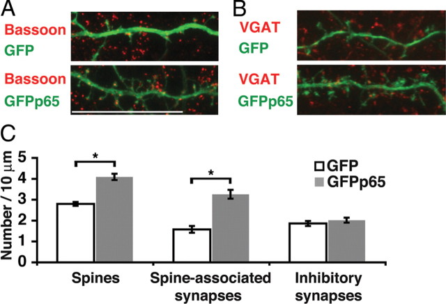
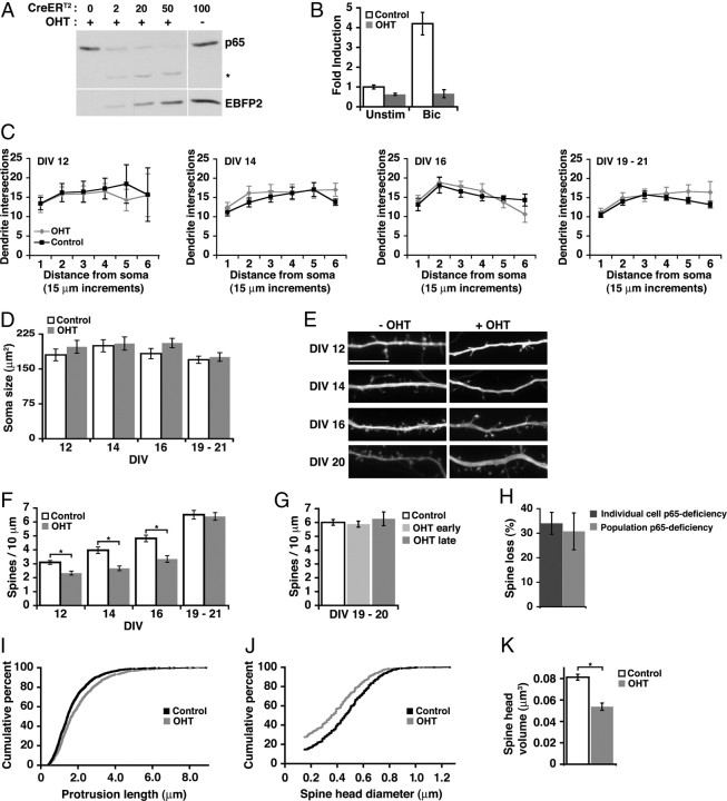
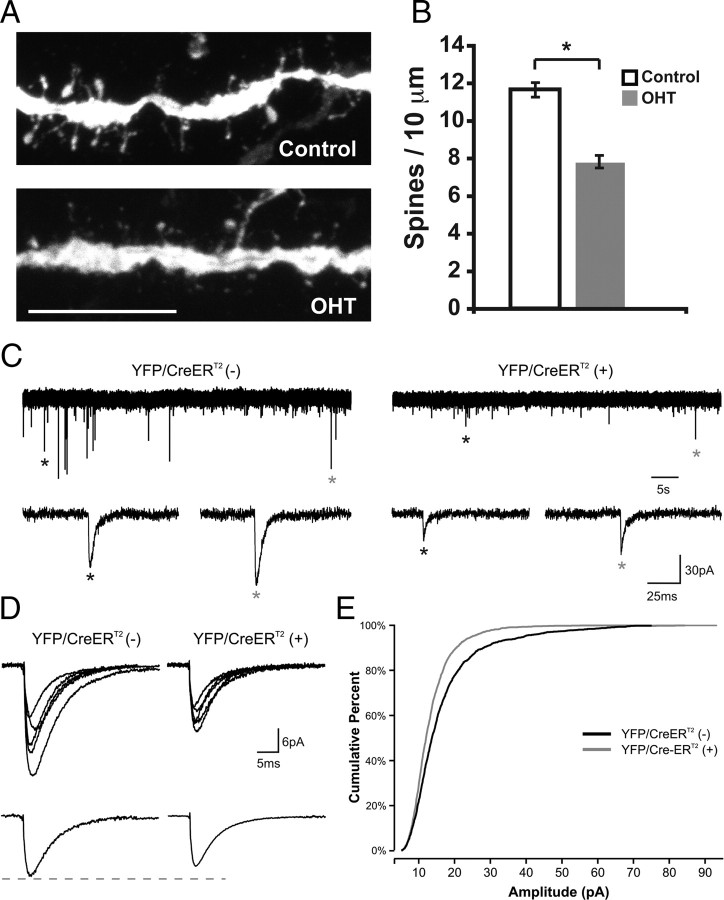
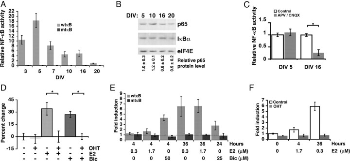
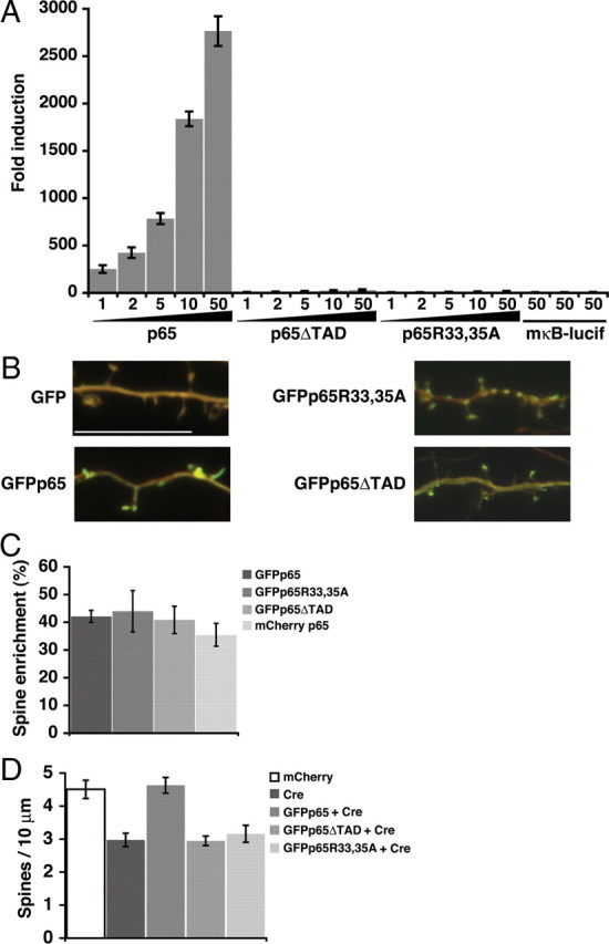
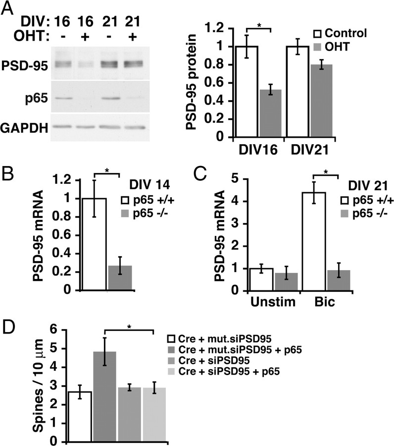
Structural plasticity of dendritic spines and synapses is a fundamental mechanism governing neuronal circuits and may form an enduring basis for information storage in the brain. We find that the p65 subunit of the nuclear factor-κB (NF-κB) transcription factor, which is required for learning and memory, controls excitatory synapse and dendritic spine formation and morphology in murine hippocampal neurons. Endogenous NF-κB activity is elevated by excitatory transmission during periods of rapid spine and synapse development. During in vitro synaptogenesis, NF-κB enhances dendritic spine and excitatory synapse density and loss of endogenous p65 decreases spine density and spine head volume. Cell-autonomous function of NF-κB within the postsynaptic neuron is sufficient to regulate the formation of both presynaptic and postsynaptic elements. During synapse development in vivo, loss of NF-κB similarly reduces spine density and also diminishes the amplitude of synaptic responses. In contrast, after developmental synaptogenesis has plateaued, endogenous NF-κB activity is low and p65 deficiency no longer attenuates basal spine density. Instead, NF-κB in mature neurons is activated by stimuli that induce demand for new synapses, including estrogen and short-term bicuculline, and is essential for upregulating spine density in response to these stimuli. p65 is enriched in dendritic spines making local protein-protein interactions possible; however, the effects of NF-κB on spine density require transcription and the NF-κB-dependent regulation of PSD-95, a critical postsynaptic component. Collectively, our data define a distinct role for NF-κB in imparting transcriptional regulation required for the induction of changes to, but not maintenance of, excitatory synapse and spine density.


Figures

Figure 1.
Figure 1.
Exogenous expression of NF-κB increases spine and excitatory synapse density. A, B, Confocal projections of dendrites and dendritic spines from DIV 16 murine hippocampal pyramidal neurons plated at low-density and immunostained for GFP and either a presynaptic marker Bassoon (A) or a marker of inhibitory presynaptic terminals VGAT (B). Neurons express either GFP or GFPp65 as indicated. Scale bar, 20 μm. C, Dendritic spine density and spine-associated predominantly excitatory synapses (colocalized Bassoon puncta) are both significantly increased (*p < 0.001) (see text) in DIV 16 pyramidal neurons expressing GFPp65. Inhibitory synapse density (VGAT staining) is not significantly different. Error bars indicate SEM.
Figure 2.
Figure 2.
Endogenous NF-κB regulates spine density and morphology during spinogenesis. A, Dose-dependent excision of p65 by CreERT2 in the presence of OHT. The indicated microliters of lentivirus coexpressing CreERT2 and EBFP2 were added to DIV 9 cultured RelAF/F hippocampal neurons, followed by 72 h of OHT exposure beginning at DIV 12; lysates were immunoblotted for p65 protein and EBFP2. The asterisk indicates p65 peptide remaining after excision. B, Loss of p65 (OHT) decreases NF-κB transcriptional activity in unstimulated cultures and prevents an increase in NF-κB-dependent transcription in stimulated conditions [bicuculline (Bic), 50 μm] as determined by luciferase reporter assay. C, D, p65 deficiency did not significantly affect dendritic arbor complexity as determined by Sholl analysis, or soma size of hippocampal pyramidal neurons at any tested DIV (ANOVA). E, Representative confocal projections of dendrites from CreERT2-expressing pyramidal neurons during maturation. Neurons were either exposed to OHT at DIV 5 to activate CreERT2 or untreated. Scale bar, 10 μm. F, Quantitation of dendritic spine density from pyramidal neurons treated as in E. Spine density was significantly decreased by loss of p65 (DIV 12, 14, 16, *p ≤ 0.005) (see text) except in mature cultures (DIV 19–21). Neurons were grown in high-density cultures. G, Delaying the time of p65 excision by exposing cultures to OHT at DIV 13–16 (OHT late), compared with DIV 5–8 (OHT early), did not uncover a role for NF-κB in regulating dendritic spines in mature cultures. H, Culture-wide p65 deficiency by viral CreERT2 introduction (Population) or focal p65-deficiency by transfected Cre (Individual) both similarly reduced dendritic spine density in DIV 16 pyramidal neurons. I, J, Cumulative distribution curves for total protrusion length (p = 4.2 × 10⁻⁹) (I) and spine diameter (J). K, Loss of p65 (OHT) significantly decreased average spine head volume in DIV 16 hippocampal pyramidal neurons (*p < 0.001) (see text) using the data in J. Error bars indicate SEM. See also supplemental Figure S1 (available at www.jneurosci.org as supplemental material).
Figure 3.
Figure 3.
Endogenous NF-κB regulates spine formation and mEPSC amplitude during synaptogenesis in vivo. A, Representative confocal projections of dendrites from YFP/CreERT2-expressing pyramidal neurons from hippocampal sections of RelAF/F × SLICK-V mice that were treated with OHT or vehicle. Scale bar, 10 μm. B, Loss of p65 (OHT) significantly decreases spine density at P12 in vivo (*p = 1.8 × 10⁻¹⁰; control, n = 35 dendrites; OHT, n = 36 dendrites; 3 brains per condition). C, Whole-cell voltage-clamp recordings in acute brain slices from RelAF/F × SLICK-V mice treated with OHT; shown are representative traces from YFP/CreERT2 (−) and YFP/CreERT2 (+) hippocampal pyramidal neurons; mEPSCs identified by asterisks are shown at expanded timescale below. D, Top, Overlay of average mEPSCs from five representative YFP/CreERT2 (−) and five representative YFP/CreER T2 (+) neurons. Bottom, Average mEPSC for all cells in each condition. E, Cumulative percentage plot of the mEPSC amplitude from all recorded neurons [n = 12 YFP/CreERT2 (−) and 15 YFP/CreER T2 (+) neurons; K-S test, p = 3.7 × 10⁻¹⁷].
Figure 4.
Figure 4.
Response to increased synaptic demand in mature neurons requires NF-κB. A, NF-κB activation was monitored by luciferase assay at time points in culture maturation preceding (DIV 3, 5), during (DIV 7, 10, 16), and at the conclusion of rapid spine and synapse formation (DIV 20). Comparison of activity from a reporter containing a wild-type NF-κB consensus binding sequence (wtκB) or a mutant binding sequence (mtκB) demonstrates specificity for NF-κB. Relative activity was plotted by normalizing fold induction to DIV 20 levels, which were the lowest observed. B, Equivalent total protein amounts from culture lysates at the indicated DIV were immunoblotted for p65, IκBα, and eIF4E (loading control); quantitated p65 levels relative to DIV 5 from two independent experiments (average ± SEM) indicate minimal changes in p65 expression. C, The effects of blockade of NMDA and AMPA receptors with APV (100 μm) and CNQX (40 μm) on NF-κB transcriptional activity at DIV 5 before rapid synapse formation and at DIV 16 (*p = 0.001) during rapid synapse formation. D, Loss of p65 after OHT addition does not decrease spine density in CreERT2-expressing DIV 20 cultured hippocampal pyramidal neurons from RelAF/F mice. Dendritic spine density in DIV 20 pyramidal neurons was increased in response to E2 (0.3 μm; 48–72 h) or bicuculline (25 μm; 24 h) by 34.1 ± 7.7 or 25.5 ± 3.6%, respectively, from untreated. Loss of p65 prevented neurons from increasing spine density in response to either E2 or bicuculline. Differences in dendritic spine density in neurons exposed to E2 or bicuculline in the presence or absence of OHT were highly significant (E2, *p = 0.003; Bic, *p = 5.5 × 10⁻⁷). E, NF-κB-dependent gene expression as determined by luciferase assay with wild-type (wtκB) or mutant (mtκB) NF-κB reporters in neurons stimulated with E2 or bicuculline for the indicated dose and duration; fold induction is relative to unstimulated. F, Loss of p65 (OHT) eliminated the induction of NF-κB activity by E2 in CreERT2-expressing hippocampal cultures from RelAF/F mice. Data are means ± SEM.
Figure 5.
Figure 5.
Requirement for NF-κB-dependent gene expression. A, Fold induction in an NF-κB luciferase reporter assay of 293T cells transfected with the indicated amount (in nanograms) of GFP-tagged wild-type p65 or transcriptionally inactive mutants (p65ΔTAD, p65R33,35A). A reporter containing a mutant NF-κB binding sequence (mκB-lucif) is not activated by transfection with 50 ng of p65, p65ΔTAD, or p65R33,35A. B, Representative confocal projections of dendrites from pyramidal neurons coexpressing mCherry and the indicated GFP or GFP-tagged p65 construct; red and green channels are overlaid to permit visualization of spine enrichment. Scale bar, 10 μm. C, Regions of interest in dendritic spines and shafts were quantitated to calculate normalized dendritic spine enrichment values and percentage enrichment relative to GFP (see Materials and Methods); wild-type p65 and mutant constructs did not significantly differ in spine enrichment. D, Expression of Cre (Cre-IRES-dsRed) in DIV 16 hippocampal pyramidal neurons resulted in a decrease in dendritic spine density that could be rescued by coexpression of GFPp65 but not by the transcriptionally inactive mutants of p65, GFPp65ΔTAD, or GFPp65R33,35A. Dendritic spine density in rescued neurons expressing GFPp65 plus Cre did not significantly differ from control neurons expressing mCherry alone (see text). Data are means ± SEM.
Figure 6.
Figure 6.
NF-κB-dependent increase in spine density involves the novel gene target, PSD-95. A, p65-deficient neurons have significantly decreased levels of PSD-95 at DIV 16 (p = 0.005), but not at DIV 21 (p = 0.09). Representative immunoblot (left) and quantitated data normalized to GAPDH (right). B, Basal expression of PSD-95 mRNA is decreased in DIV 14 p65 deficient neuronal cultures (p65−/−) compared with p65 wild-type cultures (p65+/+) (p = 0.001). C, At DIV 21, p65−/− cultures no longer have significantly lower basal PSD-95 mRNA levels, but fail to increase PSD-95 expression in response to bicuculline (Bic)-induced activation of NF-κB (p = 0.001). D, PSD-95 is required for the rescue of decreased spine density in p65-deficient neurons by p65 expression. DIV 16 pyramidal neurons from RelAF/F mice expressing Cre recombinase show a significant loss of spine density that is rescued by p65 expression in the presence of control mutant siRNA, but not in the presence of siRNA for PSD-95 (Cre/p65/mut.siPSD95, n = 8, p = 0.037; Cre/p65/siPSD95, n = 13). Data are means ± SEM. See also supplemental Figure S4 (available at www.jneurosci.org as supplemental material).

References

    1. Ahn HJ, Hernandez CM, Levenson JM, Lubin FD, Liou HC, Sweatt JD. c-Rel, an NF-kappaB family transcription factor, is required for hippocampal long-term synaptic plasticity and memory formation. Learn Mem. 2008;15:539–549. - PMC - PubMed
    1. Ai HW, Shaner NC, Cheng Z, Tsien RY, Campbell RE. Exploration of new chromophore structures leads to the identification of improved blue fluorescent proteins. Biochemistry. 2007;46:5904–5910. - PubMed
    1. Bhatt DH, Zhang S, Gan WB. Dendritic spine dynamics. Annu Rev Physiol. 2009;71:261–282. - PubMed
    1. Bonhoeffer T, Yuste R. Spine motility. Phenomenology, mechanisms, and function. Neuron. 2002;35:1019–1027. - PubMed
    1. Chen FE, Huang DB, Chen YQ, Ghosh G. Crystal structure of p50/p65 heterodimer of transcription factor NF-kappaB bound to DNA. Nature. 1998;391:410–413. - PubMed

Publication types

MeSH terms

Substances

LinkOut - more resources