Review
. 2011 Apr 6;3(77):77sr1.
doi: 10.1126/scitranslmed.3002369.

Alzheimer's disease: the challenge of the second century

Affiliations
Review

Alzheimer's disease: the challenge of the second century

David M Holtzman et al. Sci Transl Med. .

Abstract

Alzheimer's disease (AD) was first described a little more than 100 years ago. It is the most common cause of dementia with an estimated prevalence of 30 million people worldwide, a number that is expected to quadruple in 40 years. There currently is no effective treatment that delays the onset or slows the progression of AD. However, major scientific advances in the areas of genetics, biochemistry, cell biology, and neuroscience over the past 25 years have changed the way we think about AD. This review discusses some of the challenges to translating these basic molecular and cellular discoveries into clinical therapies. Current information suggests that if the disease is detected before the onset of overt symptoms, it is possible that treatments based on knowledge of underlying pathogenesis can and will be effective.


Figures
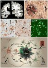
Figure 1
Figure 1. Pathology of Alzheimer’s disease (AD)
A. Postmortem brain section from an AD case (left) compared with that of a cognitively normal individual (right) reveals severe brain atrophy in AD. B. High power photomicrograph of a neuritic amyloid plaque (encircled by dashed white lines), with dystrophic neurites (small arrow) and a neurofibrillary tangle (large arrow) (photomicrograph imaged at 400X magnification; silver stain). C. Anti-Aβ antibody immunohistochemical staining of an AD brain reveals diffuse plaques containing Aβ (large arrow), compact plaques (medium size arrow) and cerebral amyloid angiopathy (smallest arrow) (photomicrograph imaged at 100X magnification). D. Anti-phospho tau antibody immunohistochemical staining reveals hyperphosphorylated tau accumulation in neuronal cell bodies (large arrow) and throughout the neuropil in neuropil threads (neuronal processes, smaller arrow, photomicrograph imaged at 200X magnification). E. Thioflavin-S stain reveals Aβ in a β-pleated sheet structure in amyloid plaques (larger arrow) and tau in a β-pleated sheet structure in neurofibrillary tangles (smaller arrow, photomicrograph imaged at 200X magnification). F. Diagram illustrating aspects of the neuropathology of AD. Aβ aggregates in the extracellular space of the brain in the form of plaques. The plaques are surrounded by astrocytes and microglial cells that secrete a variety of cytokines and other molecules including complement components, interleukin-1 (IL-1), tumor necrosis factor-α (TNFα), α-1 antichymotrypsin (ACT), α -2 macroglobulin (α2M), apolipoprotein E (apoE), and clusterin. Many neurons develop intracellular aggregates of tau in neurofibrillary tangles (NFT), and there is an increase in the amount of total tau and phosphorylated forms of tau (p-tau) into the CSF. Transport of molecules via capillaries and other blood vessels across the blood-brain barrier regulates a variety of AD-related processes. Photomicrographs in A–E courtesy of Nigel Cairns, Washington University. Panel F reprinted with permission from R. J. Perrin et al., Nature 461, 916–922 (2009).
Figure 2
Figure 2
Model of AD pathophysiology: How Aβ, tau, and other factors interact to contribute to the pathogenesis of AD. The Aβ peptide is produced from the transmembrane protein APP via cleavage by 2 enzymes, β-secretase and γ-secretase. Cleavage of APP by α-secretase prevents Aβ production. Presenilin is the active enzymatic component of the γ-secretase complex and cleaves APP at several sites within the membrane to produce Aβ peptides of different lengths such as Aβ38, Aβ40, and Aβ42. Several mutations in APP that are just outside of the Aβ region or within the coding sequence of Aβ cause forms of familial AD. Blue colored amino acids in APP/Aβ amino acid sequence represent the normal amino acids present in APP and the green colored amino acids below the normal sequence represent the amino acids that cause familial AD or CAA. Aβ is predominantly produced from APP within endosomes and a variety of molecules including the receptors LRP1 and SORL1 can influence Aβ levels. Synaptic activity also directly regulates Aβ levels. There is evidence that the Aβ aggregation is influenced by the Aβ binding molecules apoE and clusterin which likely interact in the extracellular space of the brain. A variety of molecules and processes affect Aβ clearance from the interstitial fluid (ISF) that is present in the extracellular space of the brain including neprilysin and insulin-degrading enzyme (IDE) as well as CSF and ISF bulk flow. LRP1 and RAGE appear to influence Aβ transport across the blood-brain-barrier. Aβ concentration and species (Aβ42 is more fibrillogenic) influences its aggregation. Once it aggregates into oligomers and fibrils, it can be directly toxic to cells, induce inflammation, and exacerbate the conversion of soluble tau to aggregated tau via unclear mechanisms. In addition to Aβ, a variety of factors influence tau aggregation and toxicity including tau levels, sequence, and its phosphorylation state.
Figure 3
Figure 3
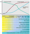
Model of the time course of biomarker changes that occur in AD in relation to the cognitive and clinical changes. Over time, in cognitively normal people that is going to develop dementia due to AD, one of the first things that occurs is the initiation of Aβ aggregation in the brain in the form of amyloid plaques. Over years, while people are still cognitively normal, amyloid plaques continue to accumulate. At some point, perhaps ~ 5 years prior to any clearcut cognitive decline, tau accumulation begins to increase in the neocortex, inflammation and oxidative stress increase, and brain network connections and metabolism begin to decline. Neuronal and synaptic loss also begins to occur as well as brain atrophy. This period when AD-type pathology is building up yet a person is cognitively normal is termed “preclinical” AD. As these changes continue to accumulate, once there is enough neuronal and synaptic dysfunction as well as cell loss, very mild dementia becomes clinically detectable. At this time, amyloid deposition has almost reached its peak. As dementia worsens to mild, moderate (mod), and severe (sev) stages, there is increasing neurofibrillary tangle formation as well as increasing neuronal and synaptic dysfunction, inflammation, cell death, and brain atrophy. Modified figure reprinted with permission from R. J. Perrin et al., Nature 461, 916–922 (2009).

References

    1. Alzheimer A. Über eine eigenartige Erkrankung der Hirnrinde. Allg Z Psychiatr. 1907;64:146–148.
    1. Barker WW, Luis CA, Kashuba A, Luis M, Harwood DG, Loewenstein D, Waters C, Jimison P, Shepherd E, Sevush S, Graff-Radford N, Newland D, Todd M, Miller B, Gold M, Heilman K, Doty L, Goodman I, Robinson B, Pearl G, Dickson D, Duara R. Relative frequencies of Alzheimer disease, Lewy body, vascular and frontotemporal dementia, and hippocampal sclerosis in the State of Florida Brain Bank. Alzheimer Dis Assoc Disord. 2002;16:203–212. - PubMed
    1. Morris JC. The Clinical Dementia Rating (CDR): current version and scoring rules. Neurology. 1993;43:2412–2414. - PubMed
    1. Petersen RC, Roberts RO, Knopman DS, Boeve BF, Geda YE, Ivnik RJ, Smith GE, Jack CR., Jr Mild cognitive impairment: ten years later. Arch Neurol. 2009;66:1447–1455. - PMC - PubMed
    1. Klunk WE, Engler H, Nordberg A, Wang Y, Blomqvist G, Holt DP, Bergstrom M, Savitcheva I, Huang GF, Estrada S, Ausen B, Debnath ML, Barletta J, Price JC, Sandell J, Lopresti BJ, Wall A, Koivisto P, Antoni G, Mathis CA, Langstrom B. Imaging brain amyloid in Alzheimer’s disease with Pittsburgh Compound-B. Ann Neurol. 2004;55:306–319. - PubMed

MeSH terms