. 2011 May 15;186(10):5638-47.
doi: 10.4049/jimmunol.1003801. Epub 2011 Apr 6.

Noncanonical K27-linked polyubiquitination of TIEG1 regulates Foxp3 expression and tumor growth

Affiliations

Noncanonical K27-linked polyubiquitination of TIEG1 regulates Foxp3 expression and tumor growth

Dong-Jun Peng et al. J Immunol. .

Abstract

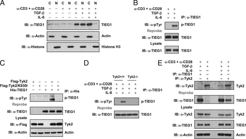
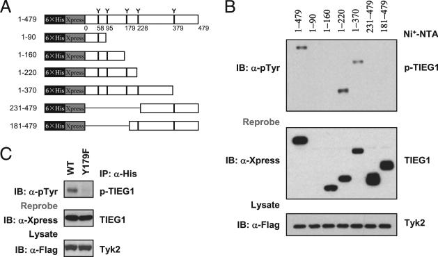
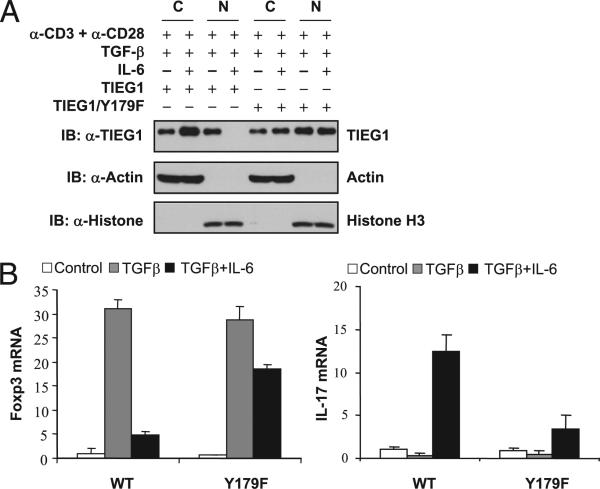
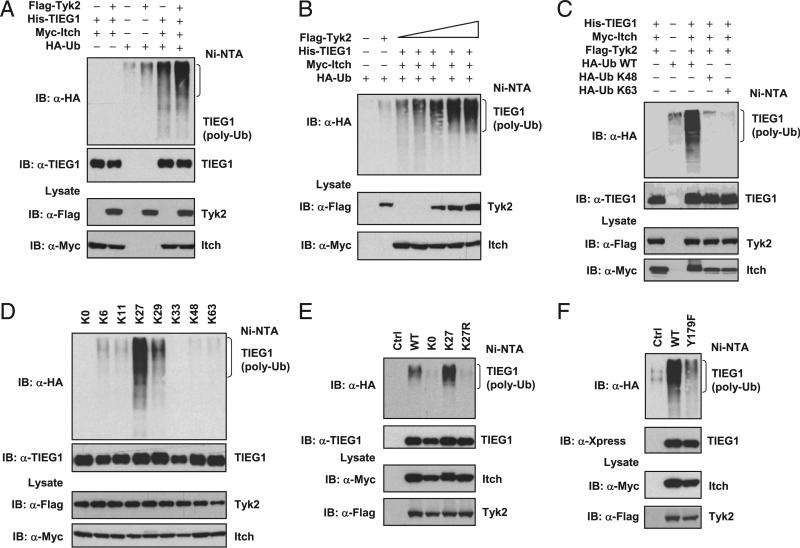
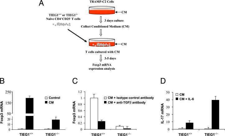
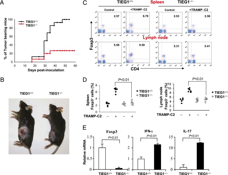
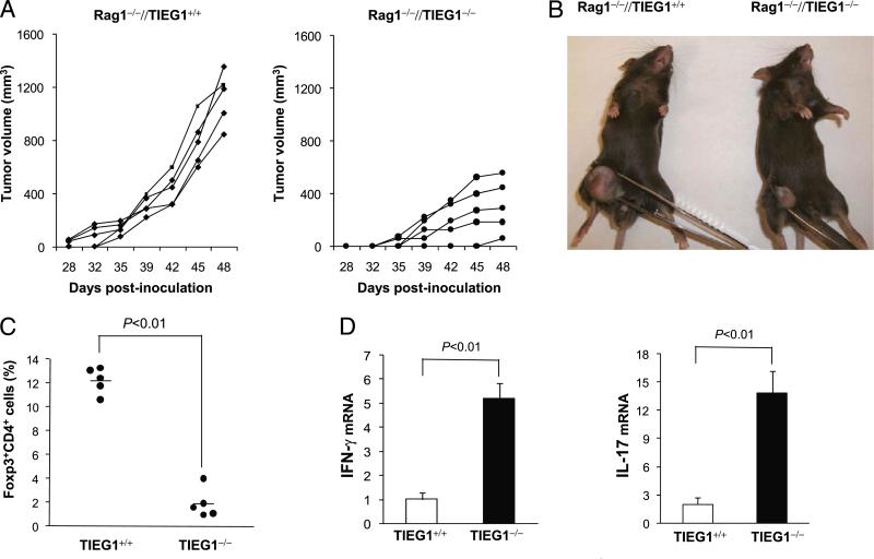
Earlier, we demonstrated the essential role of Kruppel-like transcription factor, TIEG1, in TGF-β-induced regulatory T cell (Treg) development. In this article, we demonstrate that IL-6, which promotes Th17 development, abrogated TIEG1 nuclear translocation and inhibited TGF-β-induced Treg development. Tyrosine kinase Tyk2-mediated phosphorylation of TIEG1 at Tyr179 promoted noncanonical K-27-linked polyubiquitination, which inhibited TIEG1 nuclear translocation. To test the role of TIEG1-regulated Treg/Th17 development in antitumor immunity, we analyzed TRAMP-C2 tumor growth in TIEG1(-/-) mice. The defective Treg development and elevated Th17 response resulted in enhanced immune reactivity in the tumor and inhibition of TRAMP-C2 tumor growth in TIEG1(-/-) mice. Thus, our results uncovered a novel regulatory mechanism that modulates Tregs and may regulate tumor progression.


Figures

FIGURE 1
FIGURE 1
IL-6–Tyk2 pathway-mediated phosphorylation regulates TIEG1 nuclear localization and Treg/Th17 differentiation. A, Naive CD4+CD25 T cells were stimulated with anti-CD3 and anti-CD28 in the presence of TGF-β (5 ng/ml) alone or TGF-β (5 ng/ml) plus IL-6 (20 ng/ml). The nuclear and cytoplasmic fractions were immunoblotted with anti-TIEG1 Ab. The membranes were reprobed with anti-histone H3 and anti-actin Ab. C, cytoplasmic; N, nuclear. B, CD4+CD25 T cells were treated as in A. Endogenous TIEG1 from total-cell lysates was immunoprecipitated with anti-TIEG1 Ab and immunoblotted with anti–phos-tyrosine Ab. Blots were reprobed using anti-TIEG1 Ab, p-TIEG1, or tyrosine-phosphorylated TIEG1. C, 293T cells were transfected with plasmids encoding His-TIEG1, Flag-Tyk2, or Flag-Tyk2/K930I, alone or in combination, for 24 h. TIEG1 was immunoprecipitated with anti-His Ab and immunoblotted with anti–phos-tyrosine Ab. Blot was stripped and reprobed with anti-TIEG1. The amount of Tyk2 in total-cell lysates was determined by immunoblotting. Actin was used as an equal loading control. D, Naive CD4+CD25– T cells isolated from Tyk2+/+ and Tyk2–/– mice were stimulated with anti-CD3 and anti-CD28 in the presence of TGF-β (5 ng/ml) alone or TGF-β (5 ng/ml) plus IL-6 (20 ng/ml). TIEG1 was immunoprecipitated using anti-TIEG1 Ab and immunoblotted using anti–phos-tyrosine Ab. E, TIEG1 interacts with Tyk2 in primary CD4 T cells. CD4+CD25 T cells were stimulated with TGF-β or TGF-β plus IL-6, as described above. Endogenous TIEG1 or Tyk2 was immunoprecipitated with corresponding Abs and analyzed by immunoblotting with anti-Tyk2. The same blot was stripped and reprobed with anti-TIEG1. The amount of TIEG1 and Tyk2 in total-cell extracts was determined by Western blotting. Actin was used as an equal loading control.
FIGURE 2
FIGURE 2
Tyk2 phosphorylates TIEG1 at Tyr179. A, Truncated mutants of TIEG1. Each tyrosine residue in individual truncated mutants is indicated. 6×His and Xpress represent His6 tag and Xpress tag, respectively. B, 293T cells were cotransfected with plasmids encoding Flag-Tyk2 and truncated TIEG1 for 24 h. TIEG1 was precipitated by Ni-NTA affinity purification and immunoblotted with anti–phos-tyrosine Ab (upper panel). The same blot was stripped and reprobed with anti-Xpress (middle panel). C, 293T cells were cotransfected with plasmids encoding Flag-Tyk2 and TIEG1 or TIEG1/Y179F mutant for 24 h. TIEG1 was immunoprecipitated with anti-His Ab and analyzed by immunoblotting with anti–phos-tyrosine Ab. The results shown are from one representative experiment that was conducted three times.
FIGURE 3
FIGURE 3
Tyk2-mediated phosphorylation regulates TIEG1 nuclear localization and Treg/Th17 differentiation. A, Naive TIEG1–/– CD4+ CD25 cells were reconstituted with WT or TIEG1-Y179F mutant by retroviral transduction. Nuclear/cytoplasmic localization of TIEG1 was analyzed in FACS-sorted GFP+ cells following treatment with TGF-β or TGF-β + IL-6. B, GFP+ cells were stimulated with TGF-β (Treg-inducing) and TGF-β + IL-6 (Th17-inducing) conditions for 7 d. The effect of Y179F mutation on Foxp3 versus IL-17 expression was analyzed by real-time PCR. The results shown are from one representative experiment that was conducted three times.
FIGURE 4
FIGURE 4
Tyk2-mediated phosphorylation-dependent polyubiquitination of TIEG1 is K27 linked. A, 293T cells were transfected with the indicated combination of plasmids encoding Flag-Tyk2, His-TIEG1, Myc-Itch, and HA-Ub for 48 h. Ubiquitinated TIEG1 was purified by Ni-NTA precipitation and analyzed by immunoblotting with anti-HA Ab. The expression of Tyk2 and Itch from total-cell lysates is shown (bottom two panels). B, 293T cells were transfected with plasmids encoding His-TIEG1, Myc-Itch, HA-Ub, and increased amounts of Flag-Tyk2. The ubiquitinated TIEG1 was purified by Ni-NTA precipitation and analyzed by immunoblotting with anti-HA Ab. C, 293T cells were transfected with plasmids encoding His-TIEG1, Myc-Itch, Flag-Tyk2, and HA-Ub or Ub-K48 or Ub-K63 (Ub-K48, only lysine K48 present, all other lysine mutated into arginine; Ub-K63, only lysine K63 present, all others mutated into arginine] for 40 h. Ubiquitinated TIEG1 was precipitated by Ni-NTA beads and analyzed by Western blotting with anti-HA Ab. The same blot was stripped and reprobed with anti-TIEG1. D, 293T cells were transfected with plasmids encoding His-TIEG1, Myc-Itch, Flag-Tyk2, and various lysine mutants of HA-ubiquitin (K0, all lysines are mutated into arginine; K6, K11, K27, K29, K33, K48, and K63, a single lysine present, all other lysines mutated into arginine). Ubiquitinated TIEG1 was precipitated and blotted with anti-HA Ab. E, 293T cells were transfected with Flag-Tyk2, Myc-Itch, and His-TIEG1 and HA-Ub WT, K0, K27, or K27R (K27R, only lysine K27 mutated into arginine, all other lysines present). The polyubiquitination of TIEG1 was determined by immunoblotting with anti-HA. F, 293T cells were transfected with Flag-Tyk2, Myc-Itch, HA-UbK27, His-TIEG1 WT, or His-TIEG1/Y179F mutant in 293T cells. The polyubiquitination of TIEG1 was determined by immunoblotting with anti-HA. The results shown are from one representative experiment that was conducted three times.
FIGURE 5
FIGURE 5
Defective TGF-β induced Treg development in TIEG1–/– naive CD4 T cells. A, Experimental procedures. TRAMP-C2 cells were cultured for 3 d without change of the medium. CM was collected and incubated with naive CD4+CD25 T cells isolated from spleen and lymph nodes of TIEG1+/+ and TIEG1–/– mice in the presence of anti-CD3 and anti-CD28. After 3–5 d of culture with CM, T cells were harvested for Foxp3 analysis. B, Real-time PCR analysis of Foxp3 expression. CD4+CD25 T cells were stimulated with or without CM as in A, and Foxp3 expression level was analyzed by real-time PCR. The increased folds of Foxp3 mRNA were normalized to that of actin mRNA. C, In a similar experiment to B, anti-TGF-β (15 μg/ml) or control isotype Ab was added to the cultures. Foxp3 mRNA was analyzed by real-time PCR. D, In a similar experiment to A, IL-6 was added to the T cell cultures. IL-17 mRNA was analyzed by real-time PCR.
FIGURE 6
FIGURE 6
Elevated antitumor immune response in TIEG1–/– mice. A, Kaplan–Meier curves of tumor-bearing TIEG1+/+ mice (n = 16) and TIEG1–/– mice (n = 16). Male mice were injected s.c. with TRAMP-C2 cells. Tumors were monitored three times per week. Tumor incidence in each group was calculated from days 17–60. B, Representative image of tumor-bearing TIEG1+/+ and TIEG1–/– mice. The photograph was taken at day 60 after initial inoculation. C, Foxp3+ Tregs in the spleen (upper panels) and lymph nodes (lower panels) from tumor-bearing (+TRAMP-C2) or control mice (–TRAMP-C2) were analyzed using FACS following intracellular staining using anti-Foxp3 Ab. D, CD4+Foxp3+ cell numbers in the spleen and lymph nodes of TIEG1+/+ and TIEG1–/– mice. Each symbol represents an individual mouse. E, Tumor tissues were collected from TIEG1+/+ mice or TIEG1–/– mice, and Foxp3, IFN-γ, and IL-17 expression was analyzed by real-time PCR. The results shown are from one representative experiment that was conducted three times.
FIGURE 7
FIGURE 7
Adoptive transfer of TIEG1-deficient T cells inhibits TRAMP-C2 tumor growth. Rag1–/– mice were injected s.c. with TRAMP-C2 cells on day 0. On day 3, naive CD4+CD25 (3 × 10⁶) and CD8+ T cells (3 × 10⁶) from TIEG1+/+ and TIEG1–/– mice were adoptively transferred (i.v.) into these Rag–/– mice (n = 5). A, The kinetics of tumor growth. B, Representative image of tumor-bearing Rag1–/– mice. C, Percentage of CD4+Foxp3+ cells in tumor-infiltrated cells. D, Relative mRNA levels of IFN-γ and IL-17 in tumor-infiltrated lymphocytes.

References

    1. Sakaguchi S, Powrie F. Emerging challenges in regulatory T cell function and biology. Science. 2007;317:627–629. - PubMed
    1. Chen W, Jin W, Hardegen N, Lei KJ, Li L, Marinos N, McGrady G, Wahl SM. Conversion of peripheral CD4+CD25– naive T cells to CD4+ CD25+ regulatory T cells by TGF-beta induction of transcription factor Foxp3. J. Exp. Med. 2003;198:1875–1886. - PMC - PubMed
    1. Li MO, Flavell RA. TGF-beta: a master of all T cell trades. Cell. 2008;134:392–404. - PMC - PubMed
    1. Nishikawa H, Kato T, Tawara I, Saito K, Ikeda H, Kuribayashi K, Allen PM, Schreiber RD, Sakaguchi S, Old LJ, Shiku H. Definition of target antigens for naturally occurring CD4(+) CD25(+) regulatory T cells. J. Exp. Med. 2005;201:681–686. - PMC - PubMed
    1. Nishikawa H, Kato T, Tanida K, Hiasa A, Tawara I, Ikeda H, Ikarashi Y, Wakasugi H, Kronenberg M, Nakayama T, et al. CD4+ CD25+ T cells responding to serologically defined autoantigens suppress antitumor immune responses. Proc. Natl. Acad. Sci. USA. 2003;100:10902–10906. - PMC - PubMed

Publication types

MeSH terms