. 2011 May 17;76(20):1711-9.
doi: 10.1212/WNL.0b013e31821a4439. Epub 2011 Apr 6.

Diffusion-weighted MRI hyperintensity patterns differentiate CJD from other rapid dementias

Affiliations

Diffusion-weighted MRI hyperintensity patterns differentiate CJD from other rapid dementias

P Vitali et al. Neurology. .

Abstract

Background: Diffusion-weighted imaging (DWI) and fluid-attenuated inversion recovery (FLAIR) MRI have high sensitivity and specificity for Creutzfeldt-Jakob disease (CJD). No studies, however, have demonstrated how MRI can distinguish CJD from nonprion causes of rapidly progressive dementia (npRPD). We sought to determine the diagnostic accuracy of MRI for CJD compared to a cohort of npRPD subjects.

Methods: Two neuroradiologists blinded to diagnosis assessed DWI and FLAIR images in 90 patients with npRPD (n = 29) or prion disease (sporadic CJD [sCJD], n = 48, or genetic prion disease [familial CJD, n = 6, and Gerstmann-Sträussler-Scheinker, n = 7]). Thirty-one gray matter regions per hemisphere were assessed for abnormal hyperintensities. The likelihood of CJD was assessed using our previously published criteria.

Results: Gray matter hyperintensities (DWI > FLAIR) were found in all sCJD cases, with certain regions preferentially involved, but never only in limbic regions, and rarely in the precentral gyrus. In all sCJD cases with basal ganglia or thalamic DWI hyperintensities, there was associated restricted diffusion (apparent diffusion coefficient [ADC] map). This restricted diffusion, however, was not seen in any npRPD cases, in whom isolated limbic hyperintensities (FLAIR > DWI) were common. One reader's sensitivity and specificity for sCJD was 94% and 100%, respectively, the other's was 92% and 72%. After consensus review, the readers' combined MRI sensitivity and specificity for sCJD was 96% and 93%, respectively. Familial CJD had overlapping MRI features with sCJD.

Conclusions: The pattern of FLAIR/DWI hyperintensity and restricted diffusion can differentiate sCJD from other RPDs with a high sensitivity and specificity. MRI with DWI and ADC should be included in sCJD diagnostic criteria. New sCJD MRI criteria are proposed.


Figures

Figure 1
Figure 1. Frequency of gray matter fluid-attenuated inversion recovery (FLAIR) or diffusion-weighted imaging (DWI) hyperintensities in different disease cohorts
No subject with nonprion causes of rapidly progressive dementia (npRPD) had any neocortical hyperintensity, but about 25% had predominantly limbic involvement. Almost half of Gerstmann-Sträussler-Scheinker (GSS) cases were believed to have DWI or FLAIR limbic hyperintensity. Predominant limbic hyperintensity is therefore not suggestive of sporadic Creutzfeldt-Jakob disease (sCJD), but possibly of GSS or npRPD. Familial CJD (fCJD) has an overlapping pattern of MRI abnormality with sCJD.
Figure 2
Figure 2. Three common variations of sporadic Creutzfeldt-Jakob disease presentation on MRI
(A) Neocortical (solid arrow), limbic (dashed arrow), and subcortical gray matter (dotted arrow). (B) Neocortical and limbic cortex. (C) Limbic and subcortical. Note that the diffusion-weighted imaging (DWI) shows the hyperintensities much more than the corresponding fluid-attenuated inversion recovery (FLAIR) sequences, and that DWI hyperintensities often have corresponding apparent diffusion coefficient (ADC) hypointensity. Pattern A was found in 54% of cases, pattern B in 27% of cases, and pattern C in 9% of cases. Note that the abnormalities are more readily seen on DWI than on FLAIR. ADC hypointensity, indicating restricted diffusion, corresponds to most DWI hyperintensities. ADC abnormalities are most easily identified in the basal ganglia.
Figure 3
Figure 3. Axial MRI from representative cases of familial Creutzfeldt-Jakob disease (fCJD) (A), Gerstmann-Sträussler-Scheinker (GSS) (B), and nonprion causes of rapidly progressive dementia (npRPD) (C)
(A) A fCJD case (E200K mutation) showing neocortical (solid arrow) involvement more evident in the right hemisphere, especially in the right frontal lobe, limbic involvement (dashed arrow) more evident in the right anterior cingulate and right insula and subcortical (dotted arrow) hyperintensities, greater in diffusion-weighted imaging (DWI) than in fluid-attenuated inversion recovery (FLAIR) images. Note the subcortical apparent diffusion coefficient (ADC) hypointensity in bilateral striatum. The image was read as Creutzfeldt-Jakob disease (CJD). (B) A GSS case (F198S mutation) with bilateral limbic hyperintensity in the anterior cingulate, insula, and subtle involvement in the mesiotemporal cortex, equally evident in DWI and FLAIR images. Image read as not CJD. (C) An npRPD case with limbic encephalopathy due to anti-AMPAR with anti-Sox2 antibodies and small-cell lung cancer. Note significant bilateral hyperintensity in mesiotemporal cortex (including hippocampus and amygdala), insula, and cingulate, greater on FLAIR than on DWI images. Image read as not CJD.
Figure 4
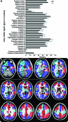
Figure 4. Frequency of gray matter hyperintensities at gyral and nuclear level in sporadic Creutzfeldt-Jakob disease (sCJD)
(A) Percent of subjects with sCJD with DWI brighter than FLAIR in each of 31 brain regions per right (black) and left (blue) hemisphere. (B) Color-coded overlay of frequency of MRI involvement in 31 brain regions per hemisphere. Light blue represents areas involved in 10%–35% of cases, dark blue 35%–50%, red 50%–65%. Neocortical regions are indicated with numbers (1–21) in the right hemisphere; limbic (22–26) and subcortical (27–31) regions are indicated in the left hemisphere.

References

    1. Geschwind MD, Shu H, Haman A, Sejvar JJ, Miller BL. Rapidly progressive dementia. Ann Neurol 2008;64:97–108 - PMC - PubMed
    1. Shiga Y, Miyazawa K, Sato S, et al. Diffusion-weighted MRI abnormalities as an early diagnostic marker for Creutzfeldt-Jakob disease. Neurology 2004;I63:443–449 - PubMed
    1. Young GS, Geschwind MD, Fischbein NJ, et al. Diffusion-weighted and fluid-attenuated inversion recovery imaging in Creutzfeldt-Jakob disease: high sensitivity and specificity for diagnosis. AJNR Am J Neuroradiol 2005;26:1551–1562 - PMC - PubMed
    1. Kallenberg K, Schulz-Schaeffer WJ, Jastrow U, et al. Creutzfeldt-Jakob disease: comparative analysis of MR imaging sequences. AJNR Am J Neuroradiol 2006;27:1459–1462 - PMC - PubMed
    1. Kretzschmar HA, Ironside JW, DeArmond SJ, Tateishi J. Diagnostic criteria for sporadic Creutzfeldt-Jakob disease. Arch Neurol 1996;53:913–920 - PubMed

Publication types

MeSH terms