Comparative Study
. 2011 Oct;60(10):1379-88.
doi: 10.1136/gut.2010.226175. Epub 2011 Apr 6.

Intracellular activation of trypsinogen in transgenic mice induces acute but not chronic pancreatitis

Affiliations
Comparative Study

Intracellular activation of trypsinogen in transgenic mice induces acute but not chronic pancreatitis

Sebastian Gaiser et al. Gut. 2011 Oct.

Abstract

Background and aims: Premature intra-acinar activation of trypsinogen is widely considered key for both the initiation of acute pancreatitis and the development of chronic pancreatitis. However, the biological consequences of intracellular trypsinogen activation have not been directly examined. To do so, a new mouse model was developed.

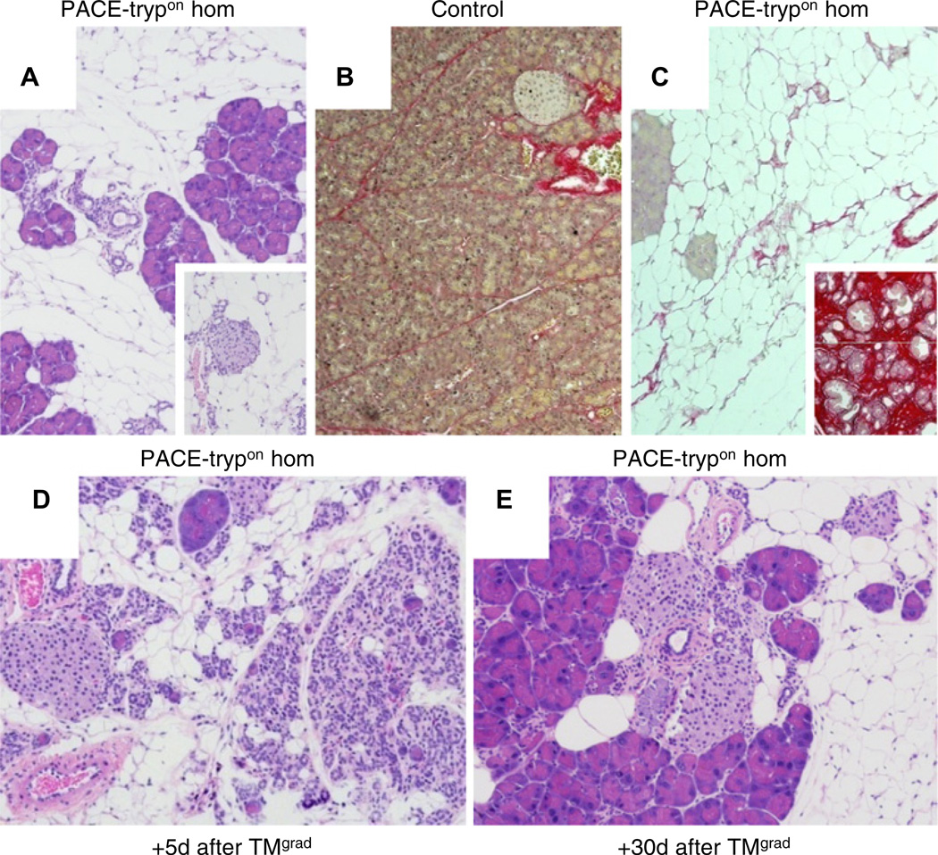
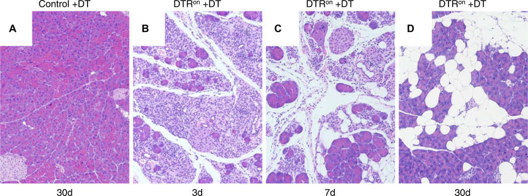
Methods: Mice were engineered to conditionally express an endogenously activated trypsinogen within pancreatic acinar cells (PACE-tryp(on)). Hallmarks of pancreatitis were determined and findings were correlated to the level (zygosity) and extent (temporal and spatial) of conditional PACE-tryp(on) expression. Furthermore, the impact of acinar cell death in PACE-tryp(on) mice was assessed and compared with a model of selective diphtheria toxin (DT)-mediated induction of acinar apoptosis.

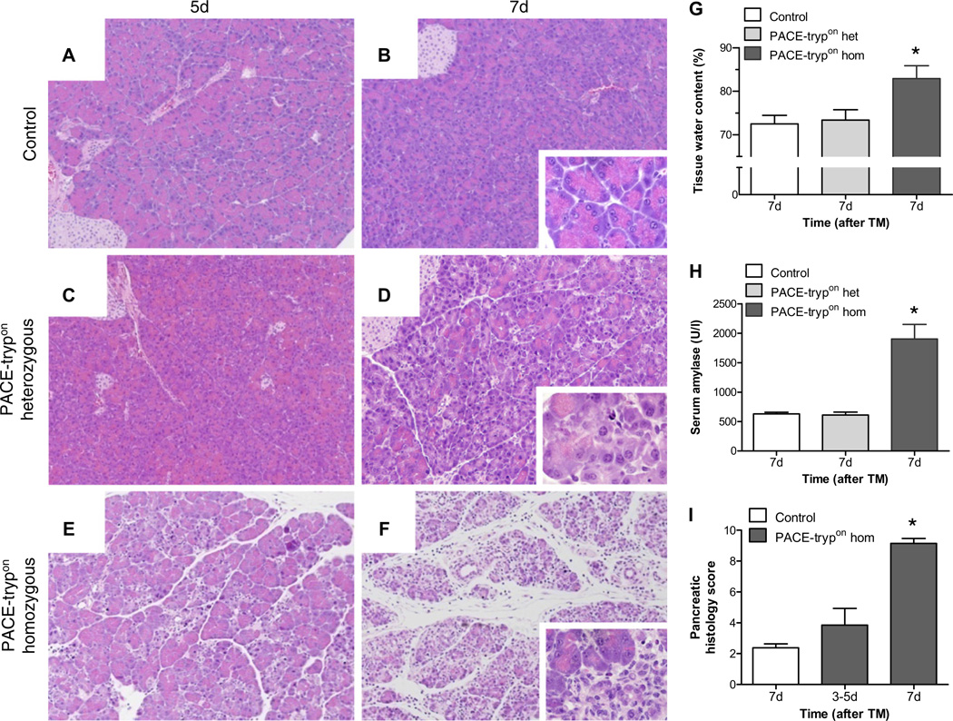
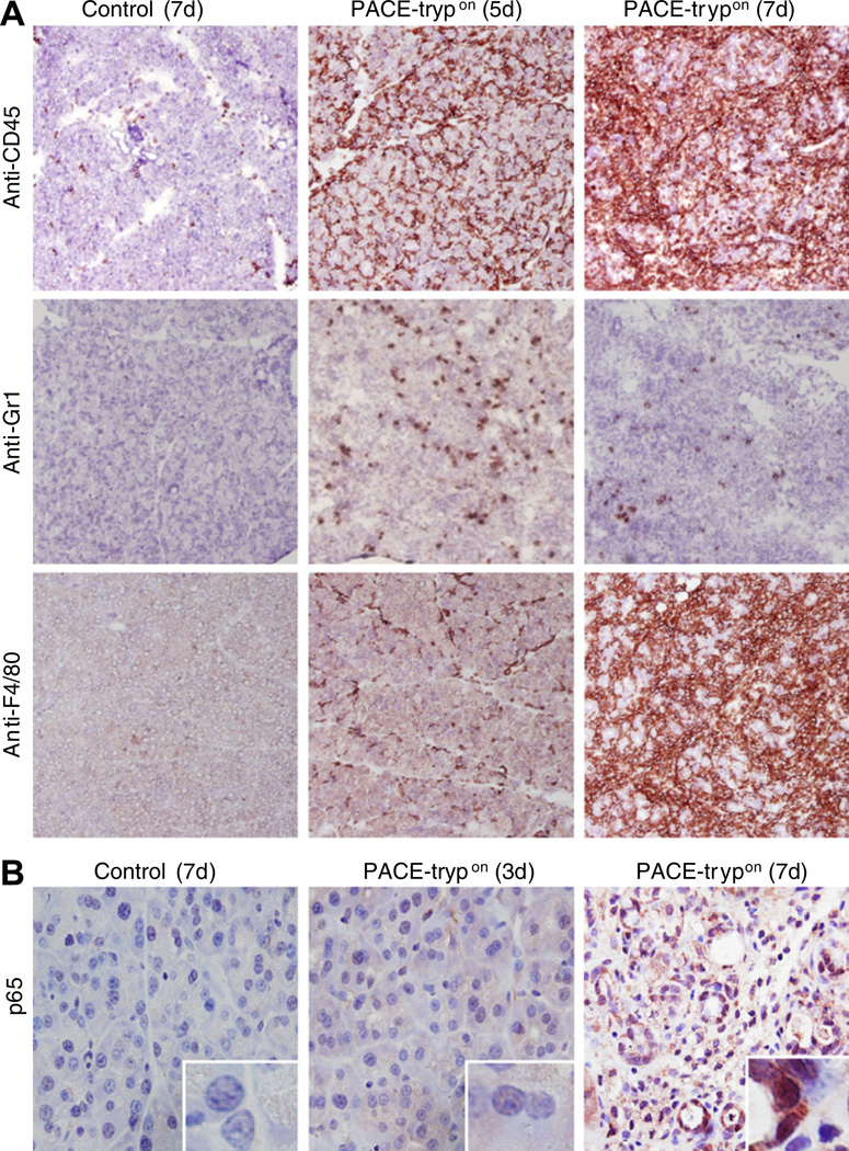
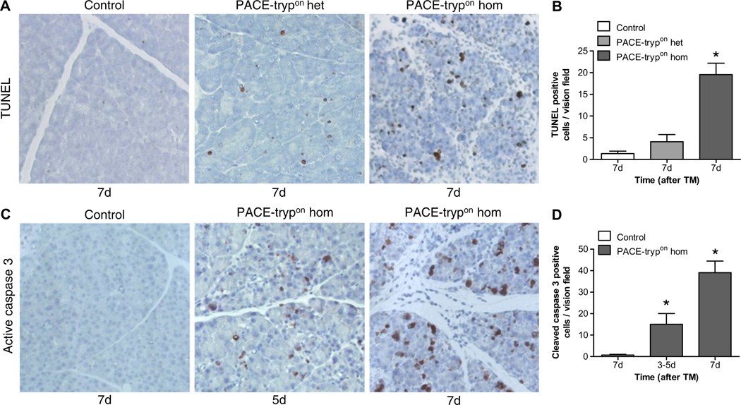
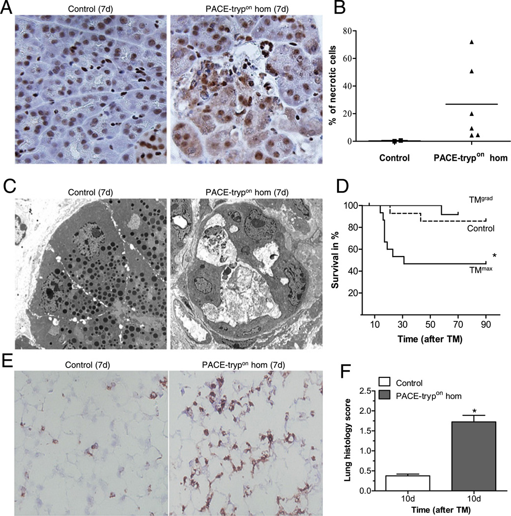
Results: Initiation of acute pancreatitis was observed with high (homozygous), but not low (heterozygous) levels of PACE-tryp(on) expression. Subtotal (maximal-rapid induction) but not limited (gradual-repetitive induction) conditional PACE-tryp(on) expression was associated with systemic complications and mortality. Rapid caspase-3 activation and apoptosis with delayed necrosis was observed, and loss of acinar cells led to replacement with fatty tissue. Chronic inflammation or fibrosis did not develop. Selective depletion of pancreatic acinar cells by apoptosis using DT evoked similar consequences.

Conclusions: Intra-acinar activation of trypsinogen is sufficient to initiate acute pancreatitis. However, the primary response to intracellular trypsin activity is rapid induction of acinar cell death via apoptosis which facilitates resolution of the acute inflammation rather than causing chronic pancreatitis. This novel model provides a powerful tool to improve our understanding of basic mechanisms occurring during pancreatitis.


Figures

Figure 1
Figure 1
Principle of PACE (paired basic amino acid-cleaving enzyme)-mediated trypsinogen activation in transgenic mice. (A) Wild-type (WT) trypsinogen is activated by enteropeptidase in the duodenum. Insertion of a PACE recognition site allows the genetically modified trypsinogen (PACE–trypsinogen) to be cleaved and activated by PACE within acinar cells. (SP, signal peptide; TAP, trypsinogen activation peptide; HA, haemagglutinin tag). (B) Induction of Cre activity in LGL-PACE-trypsinogen×CreErT double transgenic mice (PACE-trypon) removes the GFP-stop cassette. (CAG, chimeric human CMV-IE enhancer and chicken β-actin promoter; GFP, green fluorescent protein). (C) PACE–trypsinogen was expressed in PACE-trypon mice as shown in this western blot (1 week after induction). (D) Trypsin activity levels in pancreatic homogenates of PACE-trypon mice were significantly elevated 1 week after induction with tamoxifen (TM) (n=4, *p<0.05). (E) PACE–trypsinogen with an HA tag at its C-terminus was expressed in pancreatic acinar cells. The HA tag (red) and amylase (green) were visualised by immunofluorescence using anti-HA and antiamylase antibodies. Co-localization (yellow) of these two proteins indicated that active trypsin was sorted to the secretory pathway (scale bar 10 µm).
Figure 2
Figure 2
Homozygous but not heterozygous PACE-trypon mice rapidly displayed onset of acute pancreatitis. The effects of tamoxifen (TM)-induced recombination (TMmax) on control (A, B) heterozygous PACE-trypon (C, D) and homozygous PACE-trypon (E, F) mice were examined at day 5 (A, C, E) or day 7 (B, D, F) (large panels 100×, insets 600×). Some loss of zymogen granule mass was noted with heterozygotes (D), whereas rapid induction of pancreatitis with obvious damage and oedema was only observed with homozygous PACE-trypon animals (E, F). (G, H) Increased pancreatic water content verified the presence of oedema in homozygous PACE-trypon mice (G) (n=6, p<0.05) and matched elevated levels of serum amylase (H) (n=4, p<0.01). (I) Quantification of histopathological findings confirmed pancreatic injury and acute inflammation in homozygous PACE-trypon mice (control, n=8; PACE-trypon 3–5 days, n=7, 7 days, n=14; p<0.001).
Figure 3
Figure 3
Inflammatory cell infiltration and nuclear factor-κB (NF-κB) activation was evident in homozygous PACE-trypon mice. (A) Immunohistochemical staining with a pan-leucocyte marker CD45 revealed that PACE (paired basic amino acid-cleaving enzyme)–trypsinogen led to dramatically increased inflammatory cell infiltration of the pancreas over time (upper panel). Neutrophil infiltration, as indicated by Gr-1 staining, was abundant in the early phase and declined during progression of the disease (middle panel). Infiltration of mature macrophages (surface marker F4/80) occurred relatively late (lower panel) (all panels 100×). (B) NF-κB activation was detected by monitoring p65 nuclear translocation (brown staining, 400×, inserts are higher magnification of nuclei). PACE-trypon animals were sacrificed at 3 and 7 days after tamoxifen induction and localisation of p65 was conducted using an anti-p65 antibody.
Figure 4
Figure 4
Acinar cell apoptosis was prominent in PACE-trypon mice. (A) Homozygous PACE-trypon mice displayed dramatically increased numbers of TUNEL (terminal deoxynucleotidyl transferase dUTP nick end labelling)-positive cells in comparison with heterozygous animals and controls 7 days (7d) after maximal-rapid induction with tamoxifen (TMmax; 200×). (B) Quantification of apoptotic cells revealed a >15-fold increase of apoptotic cells in comparison with control (n=5, p<0.001). (C) Homozygous PACE-trypon mice showed rapid induction of caspase-3 cleavage as revealed by immunohistochemistry (200×). (D) Caspase-3-positive cells increased 10- (3–5 days) to 30-fold (7 days) in homozygous PACE-trypon mice in comparison with control (n=5, p<0.05 and p<0.001).
Figure 5
Figure 5
PACE-trypon mice displayed acinar cell necrosis and increased mortality. (A) Seven days after tamoxifen (TM) treatment, formalin-fixed pancreata were sectioned and stained with anti-high mobility group protein B1 (HMGB1) antibody. Prominent cytosolic translocation of HMGB1, a marker of necrosis, was frequently found in homozygous PACE-trypon mice but not controls (400×). (B) Counting cells with HMGB1 cytosolic translocation allowed quantitative analysis of necrotic cells in the pancreas of TM-treated control and homozygous PACE-trypon mice (7d). (C) Electron microscopy (4000×) demonstrated that control animals had no signs of acinar cell damage with normal endoplasmic reticulum and intact zymogen granules (left panel). In contrast, homozygous PACE-trypon mice (7d) showed abundant intercellular debris and severely damaged acinar cells leaking their content into the enlarged lumen (right panel). (D) Mortality in PACE-trypon mice with maximal-rapid induction with TM (TMmax) was significantly higher than those treated with gradual-repetitive induction with TM (TMgrad; control, n=15; TMgrad, n=12; TMmax, n=15; p<0.05). (E) Prominent neutrophil infiltration in the lung of homozygous PACE-trypon mice (7d) was identified by Gr-1 staining (200×). (F) Examination of lung tissue at a time point where mortality was expected to occur (10 days) revealed significantly elevated histomorphological scores (n=4, p<0.01).
Figure 6
Figure 6
PACE-trypon mice did not develop typical chronic pancreatitis. (A) Homozygous PACE-trypon mice that survived maximal-rapid induction with tamoxifen (TMmax) showed no signs of persistent acute damage, chronic inflammation or fibrosis in the long term (>10 weeks). Rather, lost acinar cells were replaced by fatty tissue. Islets of Langerhans (inset) were not affected (large panel 100×, inset 200×). (B and C) Staining for collagen with Sirius red showed little staining in control (B) or PACE-trypon mice (C). In contrast, intense staining was observed in a fibrosis model (C, inset) (all 100×). (D and E) Homozygous PACE-trypon mice that received gradual-repetitive induction with TM (TMgrad) also did not develop fibrosis. Shortly following the last TM episode (+5 days after TMgrad) acute events and ongoing recovery of pancreatic injury were observed (D). In the long term (+30 days after TMgrad), most acute events were resolved with fatty replacement of acinar cells (E) (100×).
Figure 7
Figure 7
Diphtheria toxin (DT)-induced acinar cell apoptosis caused acute inflammation and fatty replacement. (A) DT administration did not have any deleterious effects on controls (100×). (B) In contrast, DT caused acinar cell apoptosis and acute inflammation in DTRon mice by 3 days (3d; 100×). (C) Abundant inflammatory cell infiltration and fatty replacement of lost acinar cells were observed at 7 days (7d; 100×). (D) Similar to our long-term findings in PACE-trypon mice, fibrosis did not develop in these mice and lost acinar cells were replaced by adipocytes (30d) (100×).

Comment in

References

    1. Pandol SJ, Saluja AK, Imrie CW, et al. Acute pancreatitis: bench to the bedside. Gastroenterology. 2007;133(1056):e1–e25. - PubMed
    1. Whitcomb DC. Clinical practice. Acute pancreatitis. N Engl J Med. 2006;354:2142–2150. - PubMed
    1. Witt H, Apte MV, Keim V, et al. Chronic pancreatitis: challenges and advances in pathogenesis, genetics, diagnosis, and therapy. Gastroenterology. 2007;132:1557–1573. - PubMed
    1. Swaroop VS, Chari ST, Clain JE. Severe acute pancreatitis. JAMA. 2004;291:2865–2868. - PubMed
    1. Chiari H. Über die Selbstverdauung des menschlichen Pankreas. Z Heilk. 1896;17:69–96.

Publication types

MeSH terms