. 2011 Apr 7;2(4):e140.
doi: 10.1038/cddis.2011.22.

Phenethyl isothiocyanate exhibits antileukemic activity in vitro and in vivo by inactivation of Akt and activation of JNK pathways

Affiliations

Phenethyl isothiocyanate exhibits antileukemic activity in vitro and in vivo by inactivation of Akt and activation of JNK pathways

N Gao et al. Cell Death Dis. .

Abstract

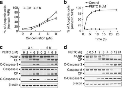
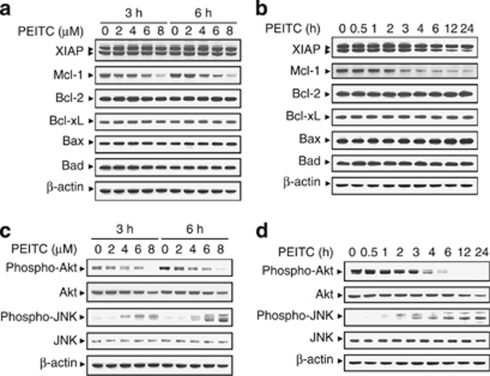
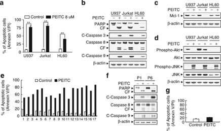
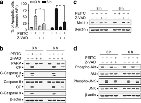
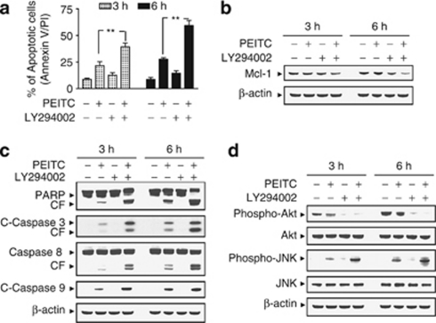
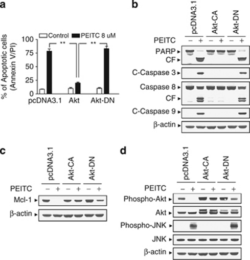
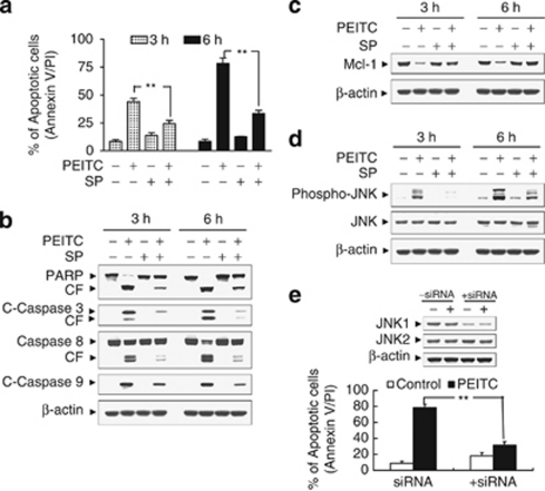
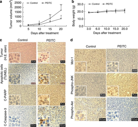
Effects of phenethyl isothiocyanate (PEITC) have been investigated in human leukemia cells (U937, Jurkat, and HL-60) as well as in primary human acute myeloid leukemia (AML) cells in relation to apoptosis and cell signaling events. Exposure of cells to PEITC resulted in pronounced increase in the activation of caspase-3, -8, -9, cleavage/degradation of PARP, and apoptosis in dose- and time-dependent manners. These events were accompanied by the caspase-independent downregulation of Mcl-1, inactivation of Akt, as well as activation of Jun N-terminal kinase (JNK). Inhibition of PI3K/Akt by LY294002 significantly enhanced PEITC-induced apoptosis. Conversely, enforced activation of Akt by a constitutively active Akt construct markedly abrogated PEITC-mediated JNK activation, Mcl-1 downregulation, caspase activation, and apoptosis, and also interruption of the JNK pathway by pharmacological or genetically (e.g., siRNA) attenuated PEITC-induced apoptosis. Finally, administration of PEITC markedly inhibited tumor growth and induced apoptosis in U937 xenograft model in association with inactivation of Akt, activation of JNK, as well as downregulation of Mcl-1. Taken together, these findings represent a novel mechanism by which agents targeting Akt/JNK/Mcl-1 pathway potentiate PEITC lethality in transformed and primary human leukemia cells and inhibitory activity of tumor growth of U937 xenograft model.


Figures

Figure 1
Figure 1
Phenethyl isothiocyanate (PEITC) significantly induces apoptosis, caspase activation, and poly(ADP-ribose) polymerase (PARP) cleavage in U937 human leukemia cells in dose- and time-dependent manners. (a) U937 cells were treated without or with various concentrations of PEITC as indicated for 3 and 6 h. (b) U937cell were treated without or with 8 μM PEITC for 1, 3, 6, 9, 12, and 24 h. Cells were stained with Annexin V/propidium iodide (PI), and apoptosis was determined using flow cytometry as described in Materials and Methods. (c) U937 cells were treated without or with various concentrations of PEITC as indicated for 3 and 6 h. (d) U937 cells were treated without or with 8 μM PEITC for 0.5, 1, 2, 3, 4, 6, 12, and 24 h. After treatment of U937 cells with the indicated PEITC concentration or the indicated time interval, total cellular extracts were prepared and subjected to western blot assay using antibodies against PARP, cleaved-caspase-3 (C-Caspase-3), caspase-8, and cleaved-caspase-9 (C-Caspase-9). Each lane was loaded with 30 μg protein. Blots were subsequently stripped and reprobed with antibody against β-actin to ensure equivalent loading and transfer. Two additional studies yielded equivalent results
Figure 2
Figure 2
Phenethyl isothiocyanate (PEITC) induces downregulation of Mcl-1, inactivation of Akt, and activation of Jun N-terminal kinase (JNK) in U937 cells. (a) U937 cells were treated without or with the indicated concentrations of PEITC for 3 and 6 h. (b) U937 cells were treated without or with 8 μM PEITC for 0.5, 1, 2, 3, 4, 6, 12, and 24 h. Total cellular extract were prepared and subjected to western blot analysis using antibody against XIAP, Mcl-1, Bcl-2, Bcl-xL, Bax, and Bad. (c) U937 cells were treated without or with the indicated concentrations of PEITC for 3 and 6 h. (d) U937 cells were treated without or with 8 μM PEITC for 0.5, 1, 2, 3, 4, 6, 12, and 24 h. Total cellular extract were prepared and subjected to western blot analysis using antibodies against cell signaling molecules, including phospho-Akt, Akt, phospho-JNK, and JNK. Each lane was loaded with 30 μg protein. Blots were subsequently stripped and reprobed with antibody against β-actin to ensure equivalent loading and transfer. Two additional studies yielded equivalent results
Figure 3
Figure 3
Phenethyl isothiocyanate (PEITC) had similar effects on apoptosis in other human leukemia cell lines as well as primary acute myeloid leukemia (AML) human leukemia cells. U937, Jurkat, and HL-60 cells were treated without or with 8 μM PEITC for 6 h, (a) after which the percentage of apoptotic cells was determined by Annexin V/propidium iodide (PI) staining and flow cytometry as described in Materials and Methods. The values obtained from Annexin V/PI assays represent the mean±standard deviation (S.D.) for three separate experiments. **Values for cells treated with PEITC were significantly increased compared with values obtained for control by Student's t-test; P<0.01. Total cellular extracts were prepared and subjected to western blot analysis using antibodies against poly(ADP-ribose) polymerase (PARP), cleaved-caspase-3 (C-Caspase-3), caspase-8, C-Caspase-9 (b), anti-apoptotic protein Mcl-1 (c), and cell signaling molecules including phospho-Akt, Akt, phospho-Jun N-terminal kinase (JNK), and JNK (d). (e) Blasts from 17 patients with AML were isolated as described in Materials and Methods. After washing and counting, isolated mononuclear cells were treated without or with 8 μM PEITC for 24 h, after which the percentage of apoptotic cells was determined by Annexin V/PI staining and flow cytometry. (f) Total cellular extracts of blasts from two AML patients (P1 and P6) were prepared and subjected to western blot analysis using antibodies against PARP, C-Caspase-3, caspase-8, and C-Caspase-9. (g) Normal peripheral blood mononuclear cells were treated without or with 8 μM PEITC for 24 h, after which the percentage of apoptotic cells was determined by Annexin V/PI staining and flow cytometry as described in Materials and Methods. The values represent the means±S.D. for three replicate determinations. For western blot analysis, each lane was loaded with 30 μg of protein; blots were subsequently stripped and reprobed with antibody against β-actin to ensure equivalent loading. Two additional studies yielded equivalent results
Figure 4
Figure 4
Phenethyl isothiocyanate (PEITC)-induced Mcl-1 downregulation, Akt inactivation, and Jun N-terminal kinase (JNK) activation were not dependent on caspase activation. U937 cells were pretreated with the caspase inhibitor benzyloxycarbonyl-Val-Ala-Asp(O-methyl)-fluoromethylketone (Z-VAD-FMK) (10 μM) for 1 h, followed by treatment with 8 μM of PEITC for 3 and 6 h. (a) Cells were stained with Annexin V/propidium iodide (PI). Apoptosis was determined using flow cytometry as described in Materials and Methods. The values obtained from Annexin V assays represent the means±standard deviation (S.D.) for three separate experiment. **Values for cells treated with PEITC and Z-VAD-FMK were significantly reduced compared with values obtained for PEITC alone by Student's t-test; P<0.01. Total protein extracts were prepared and subjected to western blot assay using antibodies against apoptosis-related proteins, including PARP, cleaved-caspase-3 (C-Caspase-3), caspase-8, and C-Caspase-9 (b); antiapoptotic protein Mcl-1 (c); as well as cell signaling proteins, including phospho-Akt, Akt, phospho-JNK, and JNK (d). For western blot analysis, each lane was loaded with 30 μg of protein, and blots were subsequently stripped and reprobed with antibody against β-actin to ensure equivalent loading
Figure 5
Figure 5
Inhibition of phosphoinositide 3-kinase (PI3K)/Akt by LY294002 (LY) enhanced phenethyl isothiocyanate (PEITC)-induced apoptosis in human leukemia cells. U937 cells were pretreated with 20 μM of LY for 1 h, followed by the addition of 4 μM of PEITC for 3 and 6 h. (a) Cells were stained with Annexin V/propidium iodide (PI), and apoptosis was determined using flow cytometry as described in Materials and Methods. The values obtained from Annexin V/PI assays represent the means±standard deviation (S.D.) for three separate experiments. **Values for cells treated with PEITC and LY in combination were significantly greater than those for cells treated with PEITC alone by Student's t-test; P<0.01. Total cellular extracts were prepared as described in Materials and Methods, and subjected to western blot analysis using antibodies against apoptosis-related proteins, including poly(ADP-ribose) polymerase (PARP), cleaved-caspase-3 (C-Caspase-3), caspase-8, and C-Caspase-9 (b), antiapoptotic protein Mcl-1 (c), and cell signaling molecules, including phospho-Akt, Akt, phospho-Jun N-terminal kinase (JNK), and JNK (d). For western blot assay, each lane was loaded with 30 μg of protein; blots were subsequently stripped and reprobed with antibody against β-actin to ensure equivalent loading. Two additional studies yielded equivalent results
Figure 6
Figure 6
Enforced activation of Akt markedly protects cells from phenethyl isothiocyanate (PEITC)-induced apoptosis. U937 cells were stably transfected with constitutively active forms of Akt (Akt-CA), dominant-negative Akt mutant (Akt-DN), and an empty vector (pcDNA3.1) as described in Materials and Methods. All cells were then treated with 8 μM of PEITC for 6 h. (a) After treatment, apoptosis was determined using Annexin V- fluorescein isothiocyanate (FITC) assay as described in Materials and Methods. **Values for Akt-CA cells treated with PEITC were significantly decreased compared with those for pcDNA3.1 and Akt-DN cells by Student's t-test; P<0.01. Total cellular extracts were prepared and subjected to western blot analysis using antibodies against apoptosis-related proteins, including poly(ADP-ribose) polymerase (PARP), cleaved-caspase-3 (C-Caspase-3), caspase-8, and C-Caspase-9 (b), antiapoptotic protein Mcl-1 (c), and cell signaling proteins, including phospho-Akt, Akt, phospho-Jun N-terminal kinase (JNK), and JNK (d). For western blot assay, each lane was loaded with 30 μg of protein; blots were subsequently stripped and reprobed with antibody against β-actin to ensure equivalent loading. Two additional studies yielded equivalent results
Figure 7
Figure 7
Effects of pharmacological and genetic interruption of Jun N-terminal kinase (JNK) on phenethyl isothiocyanate (PEITC)-induced apoptosis. U937 cells were pretreated with 10 μM of JNK inhibitor, SP600125 (SP), for 1 h, followed by the addition of 8 μM of PEITC for 3 and 6 h. (a) Cells were stained with Annexin V/PI, and apoptosis was determined using flow cytometry as described in Materials and Methods. The values obtained from Annexin V/PI assays represent the means±standard deviation (S.D.) for three separate experiments. **Values for cells treated with PEITC and SP were significantly less than those obtained for cells treated with PEITC alone by Student's t-test; P<0.01. After treatment, total cellular extracts were prepared and subjected to western blot analysis using antibodies against poly(ADP-ribose) polymerase (PARP), cleaved-caspase-3 (C-Caspase-3), caspase-8, and C-Caspase-9 (b), Mcl-1 (c), as well as cell signaling proteins, including phospho-JNK, and JNK (d). (e) U937 cells were transiently transfected with JNK1 small interfering RNA (siRNA) oligonucleotides or controls and incubated for 24 h at 37°C, after which cells were treated with 8 μM of PEITC for 6 h. Total cellular extracts were prepared and subjected to western blot analysis using antibody against JNK1 and JNK2. Apoptosis was determined using the Annexin V-FITC assay as described in Materials and Methods. **Values for cells treated with PEITC after transfection with JNK1 siRNA oligonucleotides were significantly decreased compared with those for control cells treated with PEITC by Student's t-test; P<0.01
Figure 8
Figure 8
Phenethyl isothiocyanate (PEITC) inhibits tumor growth and induces apoptosis in U937 xenograft animal model. Twenty non-obese diabetic/severe combined immunodeficiency (NOD/SCID) mice were inoculated with U937 cells (2 × 10⁶ cells per mouse, subcutaneously (s.c.)) and randomly divided into two groups (10 per group) for treatment with PEITC (50 mg/kg, intraperitoneally (i.p.), daily) or with vehicle control solvent as described in Materials and Methods. (a) Average tumor volume in vehicle control mice and mice treated with 50 mg/kg PEITC. Data are means±standard deviation (S.D.) (10 mice per group with tumors implanted on right flank of each mouse). * or **Values for tumor volume of U937 xenograft mice treated with PEITC were significantly decreased compared with vehicle control by Student's t-test; P<0.05 or P<0.01. (b) Body weight changes of mice during the 20 days of treatment. Statistical analysis of body weight changes showed no significant differences between PEITC treatment and vehicle control groups. (c) Tumors were removed from animals 20 days after the start of drug exposure. Tumors were fixed and stained with hematoxylin and eosin (H&E) stain to examine tumor cell morphology, using terminal deoxynucleotidyl transferase dUTP nick-end labeling (TUNEL) assay to determine apoptosis, and using immunohistochemistry to determine the levels of cleaved-poly(ADP-ribose) polymerase (C-PARP) and cleaved-caspase-3 (C-Caspase-3). (d) After treatment with PEITC for 20 days, tumor tissues were also sectioned and subjected to immunohistochemistry using antibodies against Mcl-1, phospho-Akt, and phospho-Jun N-terminal kinase (JNK). The sections were lightly counterstained with hematoxylin and photographed with a Scan Scope. Original magnification × 400

References

    1. Conaway CC, Yang YM, Chung FL. Isothiocyanates as cancer chemopreventive agents: their biological activities and metabolism in rodents and humans. Curr Drug Metab. 2002;3:233–255. - PubMed
    1. Stoner GD, Morrissey DT, Heur YH, Daniel EM, Galati AJ, Wagner SA. Inhibitory effects of phenethyl isothiocyanate on N-nitrosobenzylmethylamine carcinogenesis in the rat esophagus. Cancer Res. 1991;51:2063–2068. - PubMed
    1. Morse MA, Wang CX, Stoner GD, Mandal S, Conran PB, Amin SG, et al. Inhibition of 4-(methylnitrosamino)-1-(3-pyridyl)-1-butanone-induced DNA adduct formation and tumorigenicity in the lung of F344 rats by dietary phenethyl isothiocyanate. Cancer Res. 1989;49:549–553. - PubMed
    1. Morse MA, Amin SG, Hecht SS, Chung FL. Effects of aromatic isothiocyanates on tumorigenicity, O6-methylguanine formation, and metabolism of the tobacco-specific nitrosamine 4-(methylnitrosamino)-1-(3-pyridyl)-1-butanone in A/J mouse lung. Cancer Res. 1989;49:2894–2897. - PubMed
    1. Stoner GD, Morrissey DT, Heur YH, Daniel EM, Galati AJ, Wagner SA. Inhibitory effects of phenethyl isothiocyanate on N-nitrosobenzylmethylamine carcinogenesis in the rat esophagus. Cancer Res. 1991;51:2063–2068. - PubMed

Publication types

MeSH terms

Substances