

Irreducible anterior and posterior dislocation of the shoulder due to incarceration of the biceps tendon
- PMID: 21472069
- PMCID: PMC3063348
- DOI: 10.4103/0973-6042.76970
Irreducible anterior and posterior dislocation of the shoulder due to incarceration of the biceps tendon
Abstract
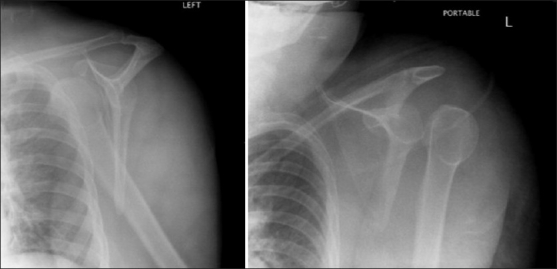
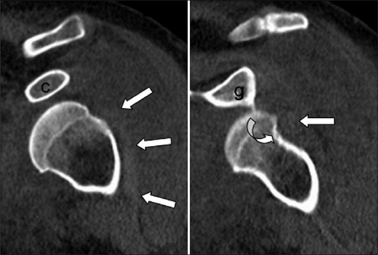
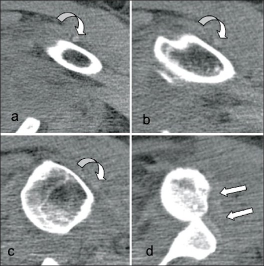
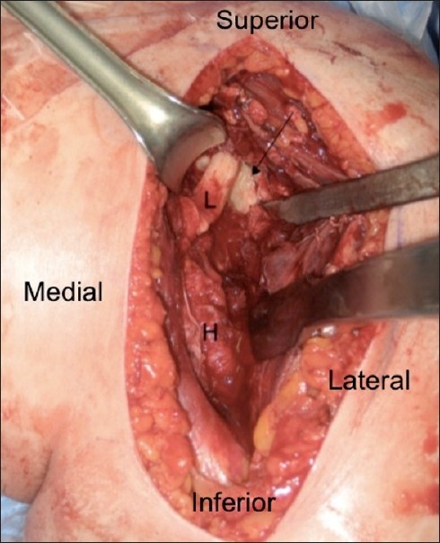
Mechanical obstacles may infrequently impede closed reduction of anterior shoulder dislocation. Imaging techniques such as arthrography, computed tomography (CT) and magnetic resonance imaging (MRI) complement conventional radiography by allowing identification of obstacles to reduction. We present a case of irreducible anterior glenohumeral dislocation resulting from an initial anterior dislocation, converted to a posterior dislocation with an attempt at reduction, then converted back to anterior dislocation with a second reduction attempt. Soft tissue obstacles to shoulder reduction should be suspected when plain films do not identify a bony fragment as the culprit. CT and MRI are useful for identifying the cause of irreducibility and for operative planning.
Keywords: Biceps tendon; computed tomography; irreducible; shoulder dislocation.
Conflict of interest statement
Figures
References
-
- Connolly S, Ritchie D, Sinopidis C, Brownson P, Aniq H. Irreducible anterior dislocation of the shoulder due to soft tissue interposition of subscapularis tendon. Skeletal Radiol. 2008;37:63–5. - PubMed
-
- Bauer T, Vuillemin A, Hardy P, Rousselin B. Posterior dislocation of the long head of the biceps tendon: A case report. J Shoulder Elbow Surg. 2005;14:557–8. - PubMed
-
- Freeland AE, Higgins RW. Anterior shoulder dislocation with posterior displacement of the long head of the biceps tendon. Arthrographic findings. A case report. Orthopedics. 1985;8:468–9. - PubMed
-
- Henderson RS. Fracture-dislocation of the shoulder with interposition of long head of biceps. J Bone Joint Surg Br. 1952;34-B:240–1. - PubMed
Publication types
LinkOut - more resources
Full Text Sources
