. 2010 Jul-Aug;3(4):581-7.
doi: 10.3892/mmr_00000301.

Imprinted tumor suppressor gene ARHI induces apoptosis correlated with changes in DNA methylation in pancreatic cancer cells

Affiliations

Imprinted tumor suppressor gene ARHI induces apoptosis correlated with changes in DNA methylation in pancreatic cancer cells

Hong Yang et al. Mol Med Rep. 2010 Jul-Aug.

Abstract

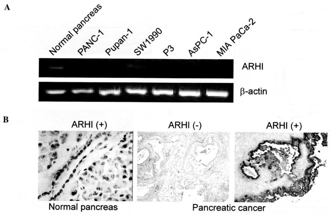
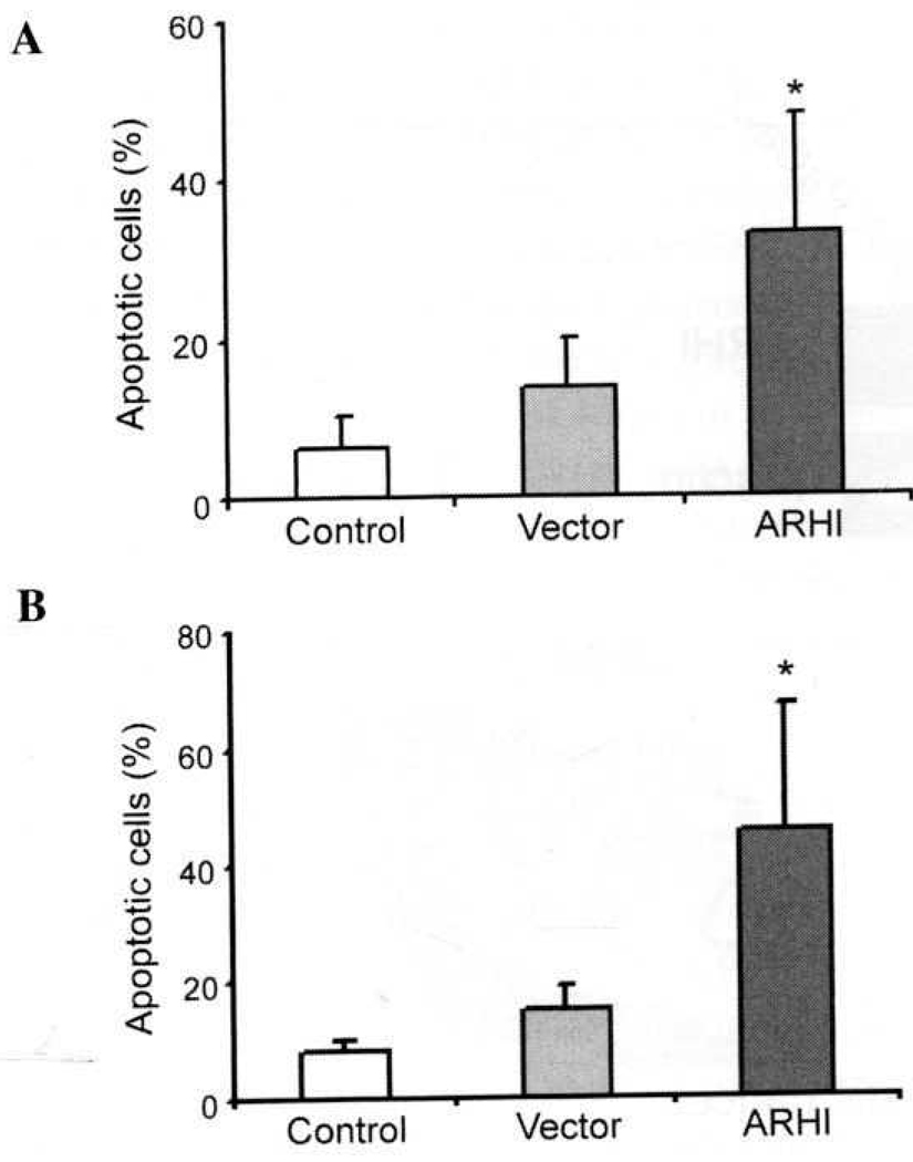
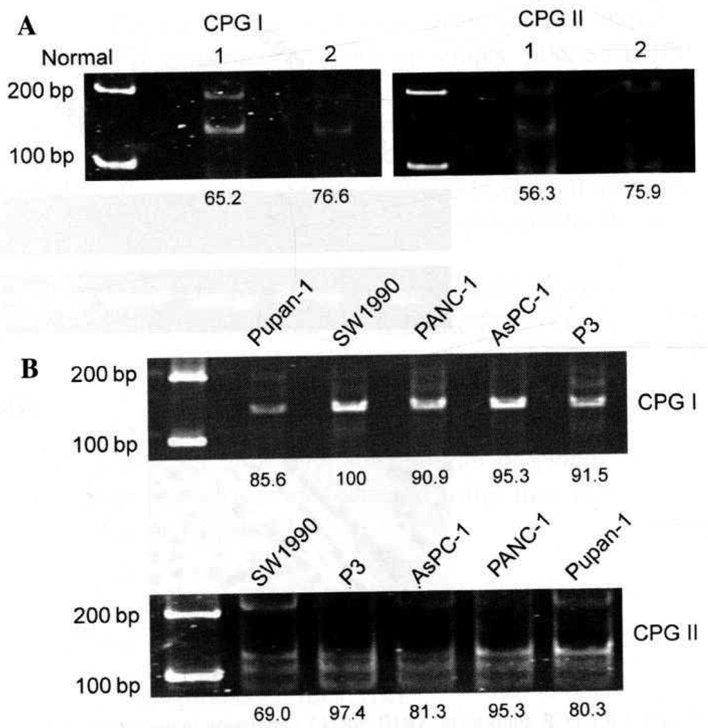
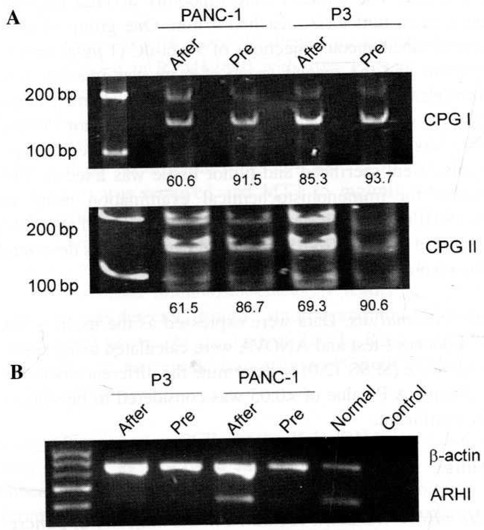
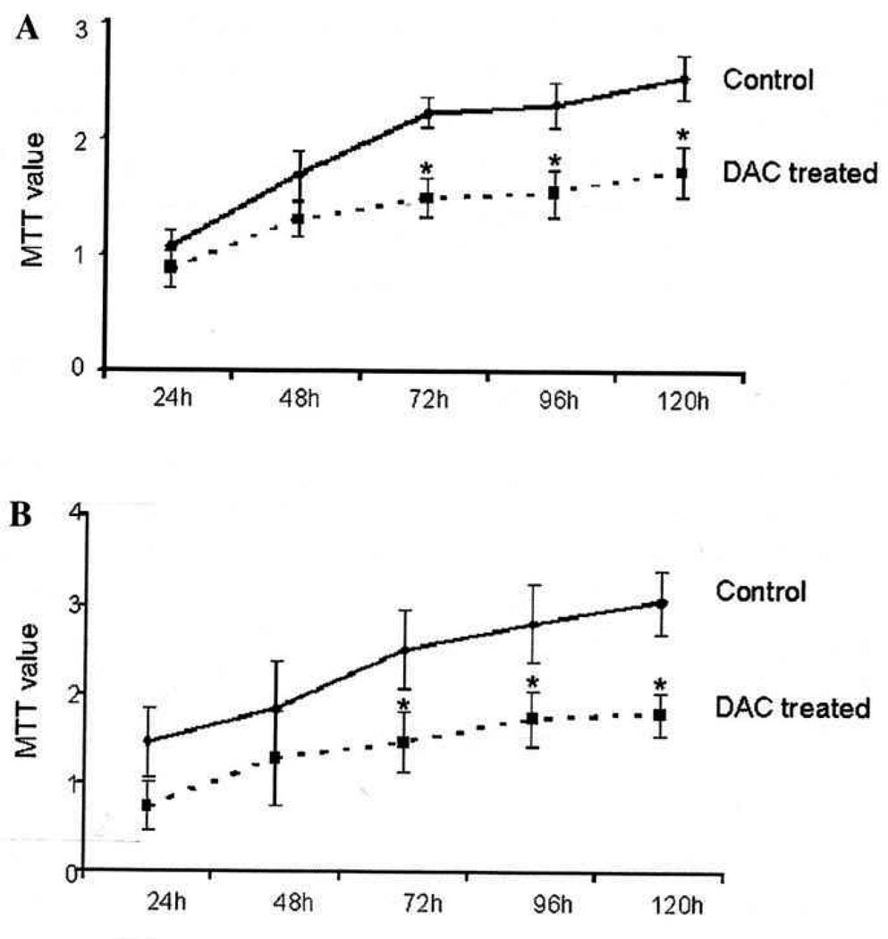
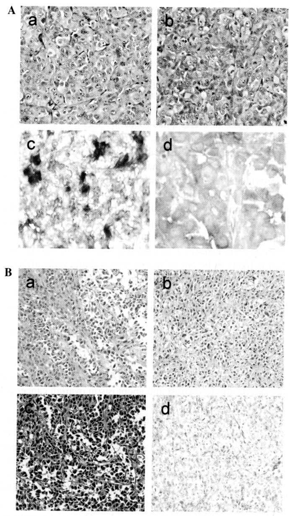
Aplesia Ras homologue member I (ARHI, DIRAS3) is a Ras-related imprinted growth inhibitory gene whose expression is down-regulated in the majority of breast and ovarian cancers. This study investigated the inhibitory function of ARHI in pancreatic cancer. Six pancreatic cancer cell lines, tumor xenografts in nude mice and 20 pancreatic cancer tissue sections were analyzed. ARHI is widely expressed in ductal and acinar cells of normal pancreatic tissue, but is down-regulated or lost in approximately 50% of pancreatic cancers. Aberrant methylation of the ARHI locus was found in five pancreatic cancer cell lines, which exhibited down-regulation or loss of ARHI expression. Hypermethylation was detected in five cell lines (5/5, 100%) at CpG island I, in two cell lines (2/5, 40%) at CpG island II and in four cell lines (4/5, 80%) at CpG island III. Re-expression of ARHI significantly inhibited the growth of pancreatic cancer cells. This inhibition was associated with the induction of apoptosis. Treatment with the demethylating agent 5-aza-2'deoxycytidine (5-aza-dC) restored ARHI mRNA expression, inhibited cell growth and induced apoptosis in PANC-1 and P3 human pancreatic cancer cells in culture. In nu/nu mice, 5-aza-dC also inhibited the growth of PANC-1 xenografts and induced apoptosis, as observed by TUNEL staining. These effects were associated with the re-expression of ARHI protein. Therefore, ARHI may serve as a growth inhibitory gene in a significant fraction of pancreatic cancers. Re-expression of ARHI significantly induced the apoptosis of pancreatic cancer cells. A demethylation agent reduced human pancreatic cancer cell line growth in conjunction with ARHI re-expression.


Figures

Figure 1
Figure 1
RT-PCR analysis of ARHI mRNA expression in pancreatic cancer cell lines and ARHI protein expression in normal pancreas and pancreatic cancer tissue by immunohistochemical staining. (A) ARHI mRNA expression was not detected in five of the six pancreatic cancer cell lines (PANC-1, P3, PuPan-1, AsPC-1 and MIA PaCa-2) by RT-PCR (above). Weak ARHI mRNA expression was observed in SW1990 cells. Normal pancreas was used as the positive control. β-actin was included as the loading control. (B) Immunohistochemical staining showed ARHI protein expression in normal pancreas and pancreatic cancer tissue (×l5). The staining occurred primarily in the cytoplasm of normal cells. By contrast, ARHI protein expression was decreased or absent in 10 of 20 (50%) pancreatic cancer tissue specimens.
Figure 2
Figure 2
Effects of ARHI re-expression on apoptosis. PANC-1 (A), MIA PaCa-2 (B) and their stable transfectants were examined for apoptosis. The apoptotic index of PANC-1 cells in the pIRES2-EGFP-ARHI group (ARHI) was significantly higher than pIRES2-EGFP (Vector) and the parental cell group (control) as determined by the Student's t-test (32.94±15.06 vs. 13.78±6.36 and 6.5±3.832%) (*P<0.05). Similar results were observed in MIA PaCa-2 cells (45.76±21.70 vs. 15.18±4.09 and 8.08±1.97%) (*P<0.05). The data shown are representative of at least five independent experiments.
Figure 3
Figure 3
Methylation status of normal pancreas tissue and aberrant methylation of ARHI CpG islands in pancreatic cancer cell lines. Two normal pancreases showed partial methylation of ARHI (CpG I and II) (A). However, hypermethylalion (both alleles) was found at CpG island I in five pancreatic cancer cell lines (100%) and in two cell lines (40%) at CpG island II (B). Methylation density (percentage of restricted vs. unrestricted fragment) was quantitated by densitometry and is shown below each lane.
Figure 4
Figure 4
Effect of 5-aza-dC on DNA methylation of ARHI and ARHI gene mRNA expression in PANC-1 and P3 cells. Treatment with 5-aza-dC decreased hypermethylation to partial methylation in P3 and PANC-1 cells (CpG islands I and II) (A). In addition, ARHI expression was restored in treated cells as compared to pre-treated cells (B).
Figure 5
Figure 5
Effect of demethylation with 5-aza-dC on PANC-1 and P3 cell growth. PANC-1 (A) and P3 cells (B) were treated with 5-aza-dC or with diluent control. The average OD value at different intervals was measured. OD values reflect the mean number of three replicate wells at different intervals from three experiments (mean ± SD, n=3). At 120 h, P3 and PANC-1 cell growth was significantly reduced (1.734±0.222 and 1.732±0.311) following treatment with 1.0 µM 5-aza-dC as compared to the diluent-treated control (2.541±0.196 and 3.051±0.346) (P<0.05).
Figure 6
Figure 6
Effect of 5-aza-dC on ARHI protein expression and cell apoptosis in nude mice tumor xenografts on Day 3 following injection with 5-aza-dC. (A) ARHI protein was partly restored in the treatment group but not in the diluent-treated control group (×20). H&E in tumor xenografts (a); positive ARHI protein expression in she same case as ‘a’ (c); H&E in tumor xenografts (b); negative ARHI protein expression in the same case as ‘b’ (d). (B) Apoptotic nuclei appeared after 5-aza-dC treatment in PANC-1 xenografts as judged by TUNEL staining (×l5). H&E staining in treatment group (a); TUNEL staining in treatment group (c); H&E staining in dilute control group (b); TUNEL staining in dilute control group (d).

Similar articles

Cited by

References

    1. Smakman N, Borel Rinkes IH, Voest EE, Kranenburg O. Control of colorectal metastasis formation by K-Ras. Biochim Biophys Acta. 2005;1756:103–114. - PubMed
    1. Kranenburg O. The KPAS oncogene: past, present, and future. Biochim Biophys Acta. 2005;1756:81–82. - PubMed
    1. Schubbert S, Shannon K, Bollag G. Hyperactive Ras in developmental disorders and cancer. Nat Rev Cancer. 2007;7:295–308. - PubMed
    1. Hingorani SR, Petricoin EF, Maitra A, et al. Preinvasive and invasive ductal pancreatic cancer and its early detection in the mouse. Cancer Cell. 2003;4:437–450. - PubMed
    1. Aguirre AJ, Bardeesy N, Sinha M, Lopez L, Tuveson DA, Horner J, Redston MS, DePinho RA. Activated Kras and Ink4a/Arf deficiency cooperate to produce metastatic pancreatic ductal adenocarcinoma. Genes Dev. 2003;17:3112–3126. - PMC - PubMed