. 2011 Aug;12(4):455-69.
doi: 10.1007/s10162-011-0267-2. Epub 2011 Apr 7.

Dynamic expression of Lgr5, a Wnt target gene, in the developing and mature mouse cochlea

Affiliations

Dynamic expression of Lgr5, a Wnt target gene, in the developing and mature mouse cochlea

Renjie Chai et al. J Assoc Res Otolaryngol. 2011 Aug.

Abstract

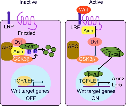
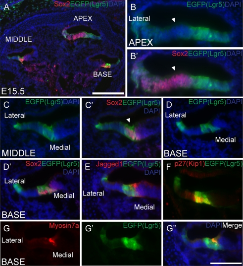
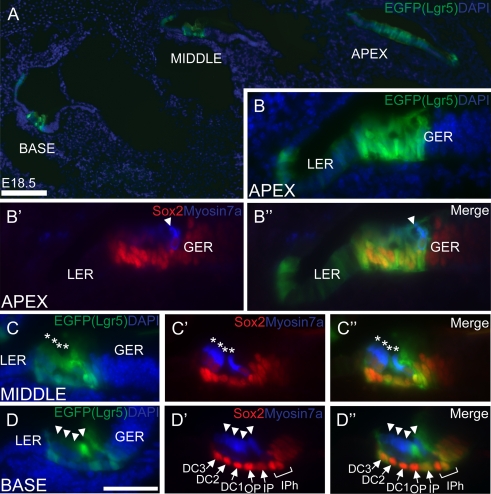
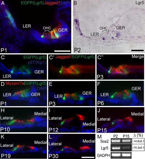
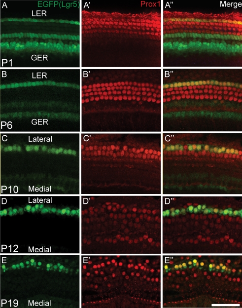
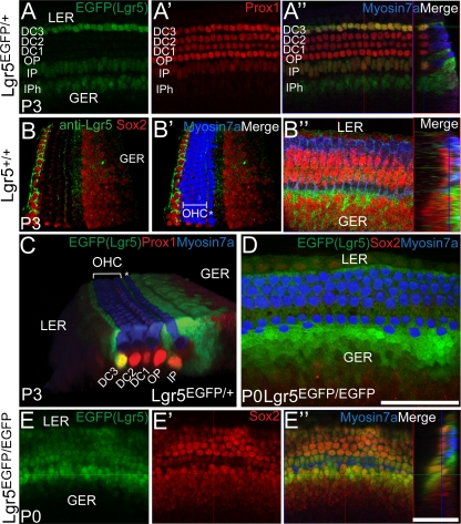
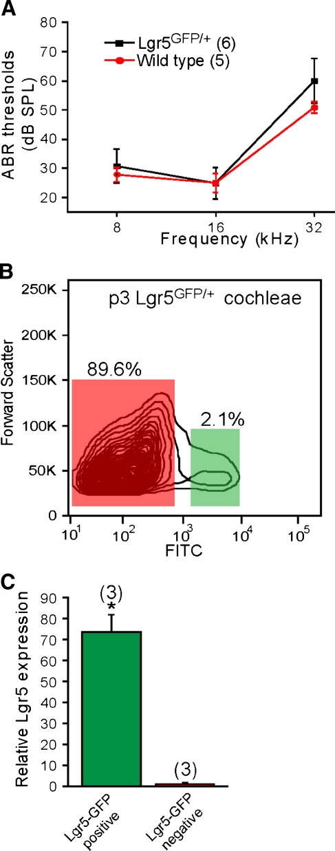
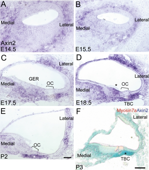
The Wnt signaling pathway is a recurring theme in tissue development and homeostasis. Its specific roles during inner ear development are just emerging, but few studies have characterized Wnt target genes. Lgr5, a member of the G protein-coupled receptor family, is a Wnt target in the gastrointestinal and integumentary systems. Although its function is unknown, its deficiency leads to perinatal lethality due to gastrointestinal distension. In this study, we used a knock-in reporter mouse to examine the spatiotemporal expression of Lgr5 in the cochlear duct during embryonic and postnatal periods. In the embryonic day 15.5 (E15.5) cochlear duct, Lgr5-EGFP is expressed in the floor epithelium and overlapped with the prosensory markers Sox2, Jagged1, and p27(Kip1). Nascent hair cells and supporting cells in the apical turn of the E18.5 cochlear duct express Lgr5-EGFP, which becomes downregulated in hair cells and subsets of supporting cells in more mature stages. In situ hybridization experiments validated the reporter expression, which gradually decreases until the second postnatal week. Only the third row of Deiters' cells expresses Lgr5-EGFP in the mature organ of Corti. Normal cochlear development was observed in Lgr5(EGFP/EGFP) and Lgr5(EGFP/+) mice, which exhibited normal auditory thresholds. The expression pattern of Lgr5 contrasts with another Wnt target gene, Axin2, a feedback inhibitor of the Wnt pathway. Robust Axin2 expression was found in cells surrounding the embryonic cochlear duct and becomes restricted to tympanic border cells below the basilar membrane in the postnatal cochlea. Both Lgr5 and Axin2 act as Wnt targets in the cochlea because purified Wnt3a promoted and Wnt antagonist suppressed their expression. Their differential expression among cell populations highlights the dynamic but complex distribution of Wnt-activated cells in and around the embryonic and postnatal cochlea.


Figures

FIG. 1
FIG. 1
Schematic for canonical Wnt signaling (modified from Reya and Clevers 2005). Wnt proteins are ligands for Frizzled receptors and low-density lipoprotein receptor-like proteins (LRP) co-receptors. In the absence of Wnt proteins, the destruction complex, which consists of Axin, adenomatous polyposis coli (APC) tumor suppressor protein, and glycogen synthase kinase 3ß (GSK3ß), phosphorylates and degrades ß-catenin, which is kept at a low cytoplasmic level. Upon activation by Wnt proteins, disheveled (Dvl) is recruited to the receptor complex and Axin is sequestered. Consequently, ß-catenin accumulates and migrates into the nucleus to bind T cell factor/lymphoid enhancer factor (TCF/LEF), which in turn activates Wnt target genes.
FIG. 2
FIG. 2
Expression of Lgr5-EGFP along the E15.5 cochlear duct floor epithelium. A At low magnification, expression of Lgr5-EGFP was restricted to floor epithelium along the cochlear duct where a differential pattern was observed along the apical–basal axis. There was no detectable expression in the roof epithelium of the cochlear duct, cells surrounding the cochlear duct, or the nascent modiolus. B, B′ In the apical turn, Lgr5-EGFP was broadly expressed along the floor epithelium and noted to be most intense in the nascent strial region and medial to the Sox2-labeled prosensory region (arrowheads). C, C′ The broad expression of Lgr5-EGFP was limited to the lateral half of the floor epithelium in the middle turn. The most intense expression was noted in the nascent strial region and in the central floor epithelium, partially overlapping with the Sox2-positive region (arrowhead). DG In the basal turn, Lgr5-EGFP was expressed in two bands of cells. The lateral band was located in the lateral sulcus, whereas the central band partially overlapped with the prosensory region as marked by Sox2 (D′), Jagged1 (E), and p27(Kip1) (F). A single row of myosin7a-positive hair cells was detected within the central band of Lgr5-EGFP-positive cells (G-G″). Scale bars, 250 μm in A and 50 μm in BG.
FIG. 3
FIG. 3
Expression of Lgr5-EGFP among distinct cell types in the E18.5 cochlear duct. A Lgr5-EGFP was expressed in a differential pattern along the apical–basal axis of the E18.5 cochlear duct. Its expression was also limited to the floor epithelium and not detected in cells surrounding the cochlear duct or nascent modiolus. BB″ Lgr5-EGFP was broadly expressed along the floor epithelium in the apical turn, with the most robust expression noted in the lateral sulcus and the prosensory region medially. The medial Lgr5-EGFP-positive domain overlapped with the Sox2-positive region and contained myosin7a-positive hair cells (arrowhead). CC″ In the middle turn, Lgr5-EGFP expression was segregated into a band in the lateral sulcus (see A) and a central band, which partially overlapped with the Sox2 marked region. Four rows of myosin7a-positive hair cells (asterisks), which co-expressed Sox2, were noted in the medial Lgr5-EGFP-positive region. Sox2 and Lgr5-EGFP were co-expressed in hair cells and supporting cells in the nascent organ of Corti. Lgr5-EGFP was expressed in the first two to three rows of cells in the lateral GER, whereas Sox2 expression spanned more medially in the GER. DD″ In the basal turn, Lgr5-EGFP was expressed in the third row of Deiters’ cells, inner pillar cells, the medial inner phalangeal cells, and the lateral GER. Its downregulation among hair cells (arrowheads) coincided with that of Sox2. DC Deiters’ cells, OP outer pillar cells, IP inner pillar cells, IPh inner phalangeal cells, LER lesser epithelial ridge, GER greater epithelial ridge. Scale bars, 250 μm in A and 25 μm in BD.
FIG. 4
FIG. 4
Lgr5-EGFP expression in specific supporting cells in the neonatal and adult organ of Corti. A, B Cryosections of Lgr5EGFP/+ cochlea (A) and wild-type cochlea processed for in situ hybridization demonstrated Lgr5 expression on the lateral cochlear wall and distinct supporting cell types. CL Cryosections of cochleae were prepared from P1-P30 Lgr5EGFP/+ transgenic mice. Lgr5-EGFP was expressed in the third row of Deiters’ cells, inner pillar cells, medial inner phalangeal cells, and the lateral GER during the first postnatal week. A decreasing gradient of Lgr5-EGFP expression was observed in the GER, with the lateral-most cells exhibiting the most robust expression. In the P3 cochlea, the cell cycle marker p27(Kip1) marked supporting cells including Hensen’s cells (C), which did not express Lgr5-EGFP and are clearly distinct from the adjacent third row of Deiters’ cells. Jagged1 was expressed on the surface membrane of supporting cells and the GER, with its expression sharply contrasted with that of Lgr5-EGFP in the P3 cochlea (C′). Between P3 and P12, the expression of Lgr5-EGFP gradually declined in the inner pillar cells, the medial row of inner phalangeal cells, and GER, but remained detectable in the third row of Deiters' cells. After P12, only the third row of Deiters’ cells in the organ of Corti expressed Lgr5-EGFP. M RT-PCR and qPCR of cochleae from P2 to P15 wild-type mice demonstrated a significant decline in Lgr5 (p < 0.001) and Sox2 expression (p < 0.05). GAPDH expression did not change significantly. Results are shown as mean ± SD. GER greater epithelial ridge, LER lesser epithelial ridge. Scale bars, 50 μm in AL.
FIG. 5
FIG. 5
Lgr5-EGFP expression becomes restricted to the third row of Deiters’ cells with age. AE Whole mounts of cochleae from P1–P30 Lgr5EGFP/+ transgenic mice were prepared and immunostained for Prox1, which marked Deiters’ cells and pillar cells. Confocal images were obtained at the level of Deiters’ cell nuclei. In the P1 cochlea, Lgr5-EGFP was expressed in the third row of Deiters’ cells, inner pillar cells, medial inner phalangeal cells, and the lateral GER. Except in the third row of Deiters’ cells, Lgr5-EGFP was downregulated in the sensory region between P1 and P12. After P12, Lgr5-EGFP was detectable only in the third row of Deiters’ cells. Scale bars, 50 μm.
FIG. 6
FIG. 6
Differential expression of Lgr5-EGFP in the Lgr5EGFP/± and Lgr5EGFP/EGFP cochleae. AA″ Confocal images of whole mount of P3 Lgr5EGFP/+ cochleae were taken at the level of Deiters’ cells nuclei. Lgr5-EGFP expression was noted in the third row of Deiters’ cells, inner pillar cells, and medial inner phalangeal cells. Reconstruction of the Z-stack images shows the segmented pattern of Lgr5-EGFP expression among these supporting cell subtypes. Prox1 marked Deiters’ and pillar cells. Myosin7a-positive hair cells did not express Lgr5-EGFP. BB″ Immunostaining for Lgr5, a transmembrane protein, in wild-type P3 cochlea demonstrated expression restricted to specific Sox2-positive supporting cell types and not myosin7a-positive hair cells. C Three-dimensional reconstruction of images illustrated that the expression of Lgr5-EGFP was restricted to subtypes of supporting cells and lateral GER. D, E Whole mounts from P0 Lgr5EGFP/EGFP cochlea showed no hair cell and supporting cell disorganization. In contrast to the heterozygous P0 cochleae where Lgr5-EGFP was detected only in the third row of Deiters’ cells, inner pillar cells, medial inner phalangeal cells, and the lateral GER, Lgr5-EGFP was also expressed in hair cells and other supporting cells in the homozygous cochleae. Confocal images captured at the level of hair cell bodies (D) and Deiters’ cell nuclei (E) of the same cochlea are shown. DC Deiters’ cells, OP outer pillar cells, IP inner pillar cells, IPh inner phalangeal cells, OHC outer hair cells, LER lesser epithelial ridge, GER greater epithelial ridge. Asterisk denotes inner hair cells in B and C. Scale bars, 50 μm.
FIG. 7
FIG. 7
Wnt signaling regulates Lgr5 expression. A, B Cochlear explants from P1 and P6 Lgr5EGFP/+ mice were incubated with purified Wnt3a (200 ng/ml) or vehicle only (1% CHAPS) for 3–4 days. Treatment with Wnt3a in the P1 cochleae did not alter the pattern of Lgr5-EGFP expression, while treatment of the P6 cochleae prevented the decline of its expression. C Treatment of cochlear explants from P3 Lgr5EGFP/+ with Fz8CRD (25 μg/ml), an inhibitor of endogenous Wnt proteins, effectively quenched Lgr5-EGFP expression. D Quantitative PCR of P3 wild-type cochlear explants treated with Wnt3a, Fz8CRD, and their respective drug vehicles for 3 days. Wnt3a significantly increased the expression of Lgr5, as well as the Wnt target genes Axin2 and Sp5 (*p < 0.01 for all three), while treatment with Fz8CRD significantly decreased their expression levels (**p < 0.001 for all three). Expression of Brn3.1 and p27(kip1) was not affected by manipulation of Wnt signals. Experiments were triplicated and expression levels were calculated using the ΔΔCT method with β-actin as the endogenous reference (Livak and Schmittgen 2001). Scale bar, 50 μm.
FIG. 8
FIG. 8
Lgr5EGFP/± mice have normal auditory function. A P30 heterozygotes and wild-type littermates were evaluated; all homozygotes died during the early postnatal periods. There were no significant differences in the auditory brainstem responses (p = 0.78) between the two groups. B, C P3 cochleae from Lgr5EGFP/+ mice were dissociated into single cells to allow for the purification of Lgr5-EGFP-positive cells via flow cytometry. Lgr5-EGFP-positive cells constituted ∼2.1% cochlear cells. Quantitative PCR demonstrated that these cells are significantly more enriched for Lgr5 expression than other cochlear cells (p < 0.001). Error bars indicate SD.
FIG. 9
FIG. 9
Axin2 expression in the embryonic and postnatal cochleae. A, B In situ hybridization detected Axin2 expression in cells surrounding the E14.5 and E15.5 cochlear duct. No significant expression was observed within the cochlear duct epithelium at these ages. C, D At E17.5 and E18.5, Axin2 was expressed in three main regions: (1) tympanic border cells (asterisks), which occupy the undersurface of the basilar membrane facing scala tympani; (2) stromal cells in the upper lateral cochlear wall; and (3) stromal cells medial to the GER which later forms the spiral limbus. Within the floor epithelium of the cochlear duct, Axin2 expression was noted in the medial GER (asterisk). E In the P2 cochlea, Axin2 expression remained robust in the tympanic border cells (asterisk) and appeared less intense in the spiral limbus and the lateral cochlear wall. F, G Cryosections of cochleae from P3 Axin2LacZ/+ transgenic mice were stained for ß-galactosidase activity. Robust expression was noted in the tympanic border cells (asterisk). Basal turns were examined in AF. TBC tympanic border cells, OC organ of Corti, GER greater epithelial ridge. Scale bars, 50 μm.

Similar articles

Cited by

References

    1. Barker N, Es JH, Kuipers J, Kujala P, Born M, Cozijnsen M, Haegebarth A, Korving J, Begthel H, Peters PJ, et al. Identification of stem cells in small intestine and colon by marker gene Lgr5. Nature. 2007;449:1003–1007. doi: 10.1038/nature06196. - DOI - PubMed
    1. Barker N, Huch M, Kujala P, Wetering M, Snippert HJ, Es JH, Sato T, Stange DE, Begthel H, Born M, et al. Lgr5(+ve) stem cells drive self-renewal in the stomach and build long-lived gastric units in vitro. Cell Stem Cell. 2010;6:25–36. doi: 10.1016/j.stem.2009.11.013. - DOI - PubMed
    1. Barolo S. Transgenic Wnt/TCF pathway reporters: all you need is Lef? Oncogene. 2006;25:7505–7511. doi: 10.1038/sj.onc.1210057. - DOI - PubMed
    1. Barolo S, Posakony JW. Three habits of highly effective signaling pathways: principles of transcriptional control by developmental cell signaling. Genes Dev. 2002;16:1167–1181. doi: 10.1101/gad.976502. - DOI - PubMed
    1. Bermingham-McDonogh O, Oesterle EC, Stone JS, Hume CR, Huynh HM, Hayashi T. Expression of Prox1 during mouse cochlear development. J Comp Neurol. 2006;496:172–186. doi: 10.1002/cne.20944. - DOI - PMC - PubMed

Publication types

MeSH terms

LinkOut - more resources