

doi: 10.1007/s13361-010-0013-8.
Epub 2011 Jan 20.
Matrix pre-coated MALDI MS targets for small molecule imaging in tissues
Affiliations
- PMID: 21472558
- PMCID: PMC4151471
- DOI: 10.1007/s13361-010-0013-8
Item in Clipboard
Matrix pre-coated MALDI MS targets for small molecule imaging in tissues
J Am Soc Mass Spectrom.
2011 Jan.
Abstract
A new sample preparation method for MALDI tissue imaging has been developed for the analysis of low molecular weight compounds that employs matrix pre-coated MALDI targets. Tissue sections need only to be transferred onto the pre-coated target before analysis for fast and easy sample preparation. Pre-coated targets have a homogenous matrix coating with uniform crystals of approximately 1-2 μm and do not require solvents that may lead to analyte delocalization within a tissue section. We report here the use of matrix pre-coated targets for imaging of lipids, peptides, and pharmaceuticals in tissues.
© American Society for Mass Spectrometry, 2011
Figures
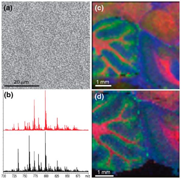
(a) SEM micrograph of DHB matrix crystals after sublimation onto a MALDI target. Scale bar = 20 μm. (b) Sum spectra from an image of a rat brain sagittal section prepared on a matrix pre-coated metal target (red) and a standard metal target that was sublimated with DHB after sectioning (black). Corresponding MALDI MS images of the brain sections prepared on a (c) matrix pre-coated target and (d) matrix post-coated target, i.e., matrix sublimated with DHB on top of tissue section. Colors correspond to ions of m/z 769.5 (SM 18:0, blue), m/z 796.5 (PC 34:2, green), and m/z 826.5 (PC 36:1, red). Images acquired at a 100-μm raster. Lipid identifications were performed by MS/MS experiments. Scale bars = 1 mm
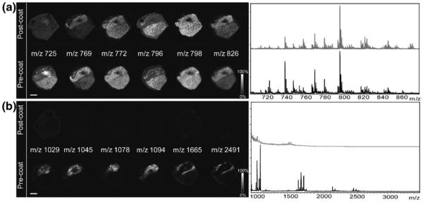
Comparison of images obtained from pituitary tissue on a DHB pre-coated target and a serial section with normal post-coating procedure each acquired with a 150-μm raster. (a) Lipid images with sum spectra shown at right. (b) Peptide images from the same tissue section with spectra shown at right. No peptides were detected when matrix was applied by sublimation only over the tissue. Scale bar = 2 mm
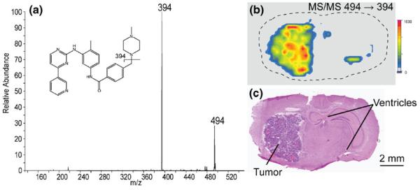
(a) MS/MS spectrum of imatinib (m/z 494). (b) The ion image of the m/z 494→394 transition of the drug showing localization to the tumor and ventricles. Image acquired with a 300-μm raster. (c) H&E stained section showing the tumor and ventricles
References
-
- Caprioli RM, Farmer TB, Gile J. Molecular imaging of biological samples: localization of peptides and proteins using MALDI-TOF MS. Anal. Chem. 1997;69(23):4751–4760. - PubMed
-
- Chaurand P, Caprioli RM. Direct profiling and imaging of peptides and proteins from mammalian cells and tissue sections by mass spectrometry. Electrophoresis. 2002;23(18):3125–3135. - PubMed
-
- Khatib-Shahidi S, Andersson M, Herman JL, Gillespie TA, Caprioli RM. Direct molecular analysis of whole-body animal tissue sections by imaging MALDI mass spectrometry. Anal. Chem. 2006;78(18):6448–6456. - PubMed
Publication types
MeSH terms
Substances
Grants and funding
LinkOut - more resources
Full Text Sources
Other Literature Sources
